Chapter 23 Tenolysis
Outline
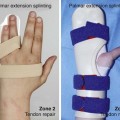
Tendon adhesions form frequently after tendon repairs and reconstructions, phalangeal fractures, and deep tissue (e.g., flexor sheath) infections.1 Multiple postoperative modalities including early active range of motion protocols have been developed to optimize tendon gliding. Aggressive therapy may be sufficient to restore full range of motion for mild adhesions.2 Alternatively, tenolysis can be a beneficial procedure for patients who have provided sufficient effort during vigorous therapy and have plateaued in their range of motion progress.3–8
One must approach these surgeries with caution, as additional surgery on a less than supremely compliant patient can lead to further edema, scaring, and worsening stiffness. Patient and physician expectations should be thoroughly discussed as it may be difficult to gain back the hand function, even if the adhesion is not extensive. Patients with severe trauma requiring multiple secondary procedures, including nerve repair, tendon grafting, capsulotomies and osteotomies, those older than 40 years, tenolysis delayed by a year, and diffuse adhesions have worse prognosis than those with isolated tendon injuries and short segments of adhesions.9
Timing of Surgery
All fractures and wounds should be healed, and chronic infections cleared. Joint contractures must be mobilized. Patient-dependent passive joint motion should be near normal prior to tenolysis.9 Occasionally, serial casting or a dynamic external fixator may be useful prior to the procedure to correct stubborn joint contractures, especially of the proximal interphalangeal joint.10
Some authors have previously recommended waiting 6 months prior to secondary tendon surgery, as stripping of the tissues surrounding the tendon may devascularize the healing tendon scar and can lead to late tendon rupture.5 Other studies show that delayed tenolysis after a year results in a decreased postoperative improvement, possibly due to a developing joint contracture.11 It has been our practice to perform tenolysis when all of the following conditions have been met: the digit and soft tissues are supple and well perfused; active motion is unacceptable to the patient; a plateau in therapy progress has been reached, with no improvement in motion over at least 4 weeks, and the patient has been cooperative with the therapy regimen. It is rare for these conditions to be met in less than 3 or even 4 months from the time of initial tendon injury or repair, but it is our opinion that tenolysis can be safely performed as soon as 3 months after the injury, providing that previously mentioned criteria have been met.
Surgical Technique
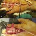
Patient involvement during the tenolysis procedure is considered important by many surgeons and is advisable whenever possible. Use of local anesthesia, with or without limited sedation, allows the patient to aid in confirmation of release of all the motion-limiting adhesions.7,12–14 Intraoperative active range of motion is the best predictor of adequate release. In addition, visualizing the expected outcome may motivate the patient to work through the tenderness of the fresh incision and new edema. While a sterile forearm tourniquet may be better tolerated than an upper arm tourniquet during the procedure, it is our preference to eschew all tourniquet use, in favor of the “wide-awake” technique advocated by Lalonde and others.13,15
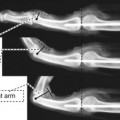
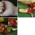
If “wide-awake” surgery is not feasible, a traction test can be used.16 The involved tendon is exposed proximal to the area of injury and retracted until digit flexion is visualized (Figure 23-1). Restrictions in movement indicate incomplete adhesion release. However, this test is not infallible; adhesions between muscle bellies may be well proximal to the zone of initial injury and surgery, especially in longer standing cases. Only “wide awake” surgery can detect such adhesions.
Related posts:

Stay updated, free articles. Join our Telegram channel

Full access? Get Clinical Tree


