Chapter 43 Tendon Gliding
The Role and Mechanical Behavior of Connective Tissues
This chapter does not appear in the print edition.
Outline
For many years, the scientific explanations concerning the natural mechanism of flexor tendon mobility in the fingers were limited to the notion of a virtual space or the existence of loose connective tissue organized in layers, but the biomechanical foundations for these theories were vague.1–3
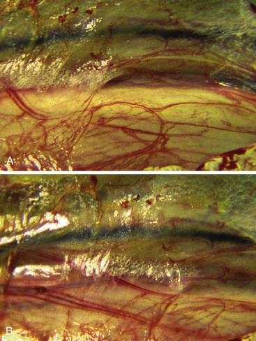
The biomechanical characteristics and histological findings of the structures around the tendons are sometimes confusing, and the roles and the definitions of the paratenon, mesotendon, peritendon, and sheaths and have largely varied among authors and their described surgical procedures4–9 (Figure 43-1).
Materials and Methods
In Vitro Study of the Paratenon
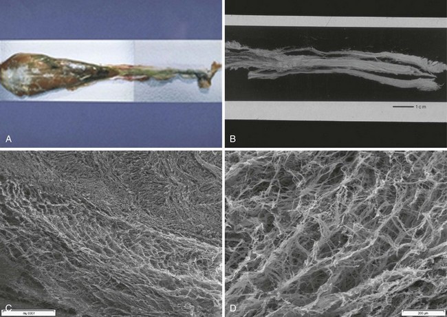
This study was carried out on 30 human upper limb biopsies of flexor digitorum superficialis (FDS) and profundus (FDP) with their surrounding sheaths, and 26 animal samples including the flexor carpi radialis from cattle where the organization is very similar to that of the human flexor profundus (Figure 43-2).
Results
In Vivo Observations
Macroscopic Observations
This suggests the existence of some sort of shock-absorbing system.
It is also clear from observing the behavior of the vessels of the common carpal sheath after revascularization and during flexion and extension, that there is apparent disorder and irregularity of shape of the microvascular network. There are surprisingly complex forms of vascular distribution, a finesse to the microvascular network, which is much more complex than a simplistic mechanistic distribution.10–14
At first sight, this is not incompatible with classical anatomical descriptions.
Ten-Fold Microscopic Examination
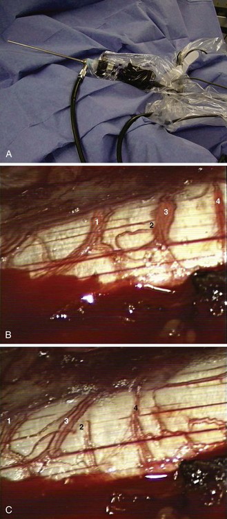
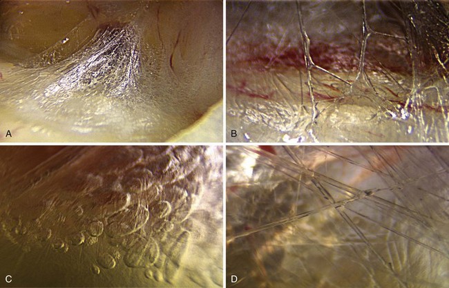
However, during flexion and extension of the tendon, tenfold microscopic examination of zones 3, 4, and 5 enabled us to observe vascular patterns in different planes of excursion and with different speeds of vessel progression due to modifications in the capillary network and depending on variations in tendon movements (Figure 43-3). Small vessels are subjected to deformation during movement but do not follow any logical or rational sequence. Some vessels progress quickly, while others move more slowly, and some overtake other vessels. The diameter of the vessel seems to be of no importance in this process. There is dynamic progression with no apparent order or proportionality.
In vivo observation has rendered this concept unacceptable partly because it is surgically impossible to define a clear field of dissection between the paratenon and the tendon (Figure 43-4).
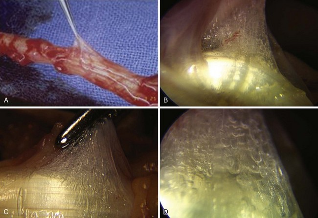
At 10-fold microscopic examination (Figure 43-5), video observation at rest showed an inaccessible microanatomical arrangement, a tissue-continuity, and a gel-like tissue surrounding the tendon. We saw a glossy structure stretching across the tendon. Within this tissue, collagen fibers can be seen framing the vessels in a random fashion. We were confronted with the notion of global dynamics and continuous matter between the tendon and the surrounding tissue, radically opposing the classical descriptions of sliding structures based on the notion of tissue stratification and a virtual space between the tissue layers. Instead, we found total histological continuity. It therefore became necessary to investigate this tissue further in order to gain better knowledge of its properties and its different roles.
At 25-Fold Magnification
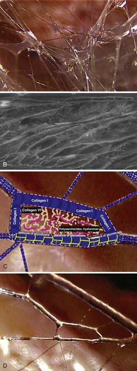
At 25-fold magnification, this glossy system consists of loose connective tissue situated between the tendon and its neighboring tissue, and is composed of intertwining multidirectional filaments creating partitions that form a new type of vacuole: three-dimensional, structured microvacuolar volumes (Figure 43-6). Apart from some adipocytes and fibroblasts, there are few cells in this multifibrillar network.
Stay updated, free articles. Join our Telegram channel

Full access? Get Clinical Tree


