Soft Tissue Coverage for Extensor Tendon Reconstruction

Chapter 35 Soft Tissue Coverage for Extensor Tendon Reconstruction


Outline


Often, extensor tendon exposure, lacerations, or tendon loss will require coverage with well-vascularized tissue to promote proper tendon healing and early mobilization. Adequate coverage also helps minimize any subsequent scarring, in an environment that is already prone to significant adhesions and contracture formation. Therefore, it behooves the reconstructive hand surgeon to provide well-vascularized tissue with either an intrinsic, locoregional, or distant free flap to provide stable soft tissue coverage. The dorsal skin of the hand has many qualities that allow flexion and extension of the digits. The increased skin laxity, pliability, and thinness also make the dorsum of the hand an excellent donor site for local flaps applicable to extensor tendon coverage. Flap selection will ultimately be dictated by the size and location of the defect, donor site availability, and recipient tissue characteristics.


Goals of this chapter will be to outline initial evaluation and management principles in the presence of exposed extensor tendons of the upper extremity following injury. Principles of flap selection and use will be discussed to provide stable coverage and expedite rehabilitation and maximize function.


Wound Débridement


Early radical débridement, of all nonviable tissue under tourniquet, control is pivotal prior to any attempt at locoregional or free flap soft tissue reconstruction. The critical step involves débridement of all marginally and questionably viable tissue to convert a contaminated wound into a clean wound and thus minimize risks of subsequent infection. This also allows a clear evaluation of all injured and missing structures. Early aggressive débridement should be performed over traditional serial débridement whenever possible. Serial débridement can delay final wound closure and make assessment of tissue viability difficult, and trigger a cascade of additional tissue loss from desiccation due to prolonged dressing changes. Early débridement and reconstruction also help to achieve the ultimate goal of early mobilization and return to function, which is critical in the upper extremity.


The general assessment is done, ideally, in the operating room under tourniquet control. The upper extremity is exsanguinated and a tourniquet is elevated and used to provide a blood-free field, so as to better expose and identify potentially devitalized tissue. All devitalized tissue is then débrided and the wound is extirpated very much like a tumor from the periphery towards the center and within well-defined natural tissue planes. The wound bed is resected entirely thus converting it from a contaminated to a noncontaminated wound. Following wound débridement, and once the extensor tendons have been either grafted or repaired, healthy coverage can be provided with either an intrinsic, locoregional, or free flap, as needed. A major prerequisite is adequate wound débridement to minimize any risk of infection, wound breakdown, and flap loss. Any initial wound that cannot be débrided thoroughly is best débrided serially after 24 or 48 hours. A negative pressure dressing can be applied temporarily using a white sponge over exposed extensor tendons so as to minimize any risk of desiccation. Note that negative pressure therapy should be used sparingly and only as a temporizing measure prior to definitive closure, which should be performed ideally within the first week. The upper extremity is very sensitive to the timing of reconstruction, and any prolonged immobilization will have a definite negative impact on final range of motion due to progressive stiffness, joint contracture, and swelling. Early coverage allows patients to enter an aggressive rehabilitation program to maximize their range of motion.


Once the wound has been properly débrided, the tourniquet is released and all nonbleeding and nonviable tissue is reexcised. Wound edges are inspected for bright red bleeding, and the wound is irrigated with a bulb syringe only. Pulse lavage is not used and can lead to extensor tendon friability and additional soft tissue trauma. Therefore, we only use triple antibiotic saline with bulb irrigation.


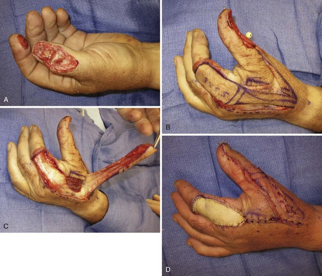
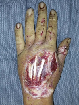
Choice of extensor tendon coverage, working from distal to proximal, will be based on the level of injury as well as the availability of nontraumatized soft tissue in the proximity of the wound. Clearly, replacing like with like is the ideal condition. All defects that are replaced with local tissue of similar color, texture, pliability, and sensation will provide a better outcome than non loco-regional flaps. The resulting defect, following radical débridement, will also be much larger than the initial wound size. Knowing that stable soft tissue coverage can be provided regardless of defect size should be impetus for providing aggressive initial débridement. This not only serves to expedite reconstruction but also helps minimize any risk of infection due to the presence of residual necrotic tissue (Figure 35-1).


Soft Tissue Coverage and Reconstruction


Reverse Cross-Finger Flap


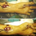
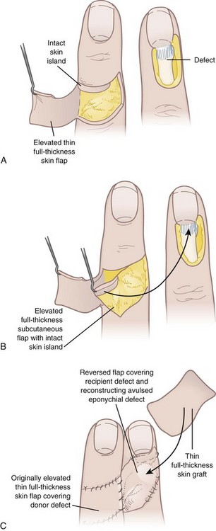
Extensor tendon exposure in zones 1 through 4 can be covered with reverse cross-finger flaps. The reverse cross-finger flap, first described by Pakiam in 1978, is an excellent local flap for soft tissue coverage of exposed extensor tendons in the dorsal digit. Its vascular supply is provided by the dorsal digital branches of the digital arteries, as well as small subcutaneous veins and venae comitantes from the dorsal digital branches. Subcutaneous veins from the cross-finger flap can also be used as venous flow through veins for replants to bridge both recipient and donor veins. The reverse cross-finger flap is then covered with either a split- or full-thickness skin graft for definitive coverage. The flap is elevated as follows: The adjacent donor digit is used for a cross-finger flap. The mid-axial skin incision is made proximal to the defect, and the skin flap is elevated just above the subcutaneous tissue in a direction that is opposite the side of the defect. The adipofascial flap is then harvested down through paratenon in the opposite direction of the elevation of the overlying skin flap. This is taken from mid-axial line to mid-axial line. Care is taken to not damage any of the dorsal branches of the digital artery when harvesting this flap. The flap is then turned over to cover the adjacent digit dorsal defect and provide good coverage for the exposed extensor tendon. The cross-finger adipofascial flap is covered with a split-thickness skin graft and the donor site is closed primarily with its hinged skin flap. The hand is immobilized for 2 to 3 weeks, preferably 3 weeks, and the division and inset of the flap is performed after 2 to 3 weeks, with revisions to the donor or recipient site as needed. The patient is splinted in a position of function for 3 weeks to minimize any contractures. In compliant patients, the patient can begin light active range of motion after the surgery but needs to be very compliant (Figure 35-2).


First Dorsal Metacarpal Artery Flap


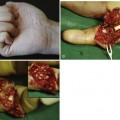
The first dorsal metacarpal artery flap (FDMA flap, i.e., kite flap), described by Foucher and Braun,1 provides two major applications in hand reconstruction: (1) dorsal hand wound coverage and (2) thumb reconstruction.


In Foucher’s anatomical study of 30 injected cadaveric hands, the FDMA originated from the radial artery in 28 of 30 specimens and from the dorsalis superficialis antebrachialis artery in 2 of 30 cases. From there, it courses distal to the extensor pollicis longus tendon, and proximal to the radial artery’s entry between both heads of the first dorsal interosseous (DIO) muscle. The FDMA travels parallel to the dorsal surface of the second metacarpal, and superficial to the first DIO muscle fascia, with some fibers occasionally covering the vessel. The FDMA then continues distally and anastomoses at the level of the metacarpal neck with dorsal perforating branches from the palmar metacarpal arteries of the deep palmar arch. These perforator branches form the basis of the reverse flow FDMA island flap. They anastomose with three different arterial systems: (1) distally with the dorsal branches of the proper palmar digital arteries, (2) proximally with branches of the DMA, and (3) laterally with adjacent DMA perforating branches (Figure 35-3).


Mar 5, 2016 | Posted by in Hand surgery | Comments Off on Soft Tissue Coverage for Extensor Tendon Reconstruction

Full access? Get Clinical Tree

Get Clinical Tree app for offline access