The introduction of perforator flaps by Koshima and Soeda in 1989 was met with much animosity in the surgical community. The flaps challenged conventional teaching and were often branded as being unsafe. Surgeries using perforator flaps are now routinely practiced all over the world, with increasing emphasis on minimizing donor site morbidity, and perforator flaps are becoming the current gold standard. The simple principles and techniques of perforator dissection can be applied to all perforator flaps, provided the surgeon has an intimate knowledge of the regional anatomy. Thus, virtually any piece of skin can be harvested as long as it incorporates a feeding vessel. This article highlights the essential techniques in planning and raising perforator flaps and the common pitfalls to be avoided.
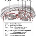
In 1989, Koshima and Soeda described a free flap that is based solely on a perforating vessel, which emanated from the deep inferior epigastric artery. Thus, it became apparent that flaps could be supplied by vessels previously dismissed as being too small to be considered. This discovery represented an evolution in flap surgery and the foundation on which a new genre of flaps was discovered.
The principal advantage of perforator flaps is that they spare deeper structures and thus limit functional morbidity. The size of the skin paddle that can be raised is, to a certain extent, related to the size of the perforator. Thus, inclusion of muscle has no influence on flap perfusion and it should be spared whenever possible. The disadvantages of perforator flaps lie in their technical demands and the resultant potential increase in operative time. The dissection of the flap requires meticulous attention to detail and a high degree of flexibility of the operative plan according to the position, size, and presence of perforating vessels. However, the somewhat unpredictable nature of the vessels can be partly addressed by accurate preoperative imaging.
Preoperative planning
Flap Planning
Accurate preoperative planning is an essential component of perforator flap surgery. Preoperative planning begins with delineation of the anatomic defect to replace “like with like”. Both the dimensions and constituent parts of the defect need to be considered. Practically, this consideration can be divided into the surface area of the skin paddle, the thickness and consistency of the subcutaneous layer, the total flap volume, and any specialized structures. If a complex reconstruction is necessary, the flap may need to incorporate specific anatomic components, such as bone, fascia, muscle, and nerve. The use of a perforator flap in these circumstances allows the skin flap to be mobilized from the other constituents to give flexibility in its placement. In this instance, the bone, muscle, and other such parts can be placed to permit a functional reconstruction, with the skin paddle draped to provide optimum contour. These have been termed chimeric flaps and are discussed later.
Having considered what needs to be replaced, the surgeon must give consideration to the most appropriate donor site and method of flap transfer (pedicled or free). For practical purposes, it is also preferable to select a flap that does not require position changes and facilitates a 2-team approach.
Although the technical aspects of flap dissection are similar for pedicled and free flaps, there are essential differences in flap planning and raising. The epicenter of a free perforator flap must lie at the level of the feeding vessel to ensure adequate tissue perfusion to all zones.
Local perforator flaps offer limited flap movement around the perforator, which represents the central axis of rotation. The extent of the flap movement depends on the tissue elasticity and perforator vessel length. The latter can be increased by following the perforator into the fascia or muscle. Several studies have shown that longer pedicles are less sensitive to twisting forces, because the length of a vessel is inversely proportional to the critical angle of twisting. Flaps that include only 1 perforator are likely to have greater flap mobility and can be mobilized in a propeller fashion, 180° counterclockwise or clockwise, without compromising perfusion. Flaps with multiple perforators are more suitable for rotational or advancement maneuvers, depending on the number of vessels preserved. If more than 1 perforator is included, then the vessels must be in close proximity to each other and dissected for a sufficient distance.
The position of the perforator selected depends on the planned movement of the flap. In a propeller flap, the perforator closest to the defect is chosen to allow the flap to pivot on the vessel and thus increase the potential coverage of the defect. For advancement, transposition, and rotation flaps, perforators furthest from the defect are selected because this provides the longest possible pedicle thereby giving the flap a large arc of motion. Regardless of the type of flap movement, any stretching of the perforator vessels should be avoided to minimize the risk of vascular complication.
Perforator flaps can also be based on a known named perforator (in a similar manner to standard musculocutaneous or fasciocutaneous flaps) or they can be designed freestyle on a random unnamed perforator of sufficient caliber.
Freestyle flaps can be considered as a form of reverse planning. The required characteristics of the skin flap determine its anatomic location, and the perforating vessels are selected as a secondary consideration. The benefits of this method lie in its flexibility in assuring that the most suitable flap is raised. However, the surgeon must be competent in locating the perforators with an ultrasound or duplex Doppler probe and must possess an excellent anatomic knowledge of the region. These flaps can be technically demanding and often require dissection and anastomosis of vessels that are considerably smaller than standard perforator flaps.
Flap design modifications and refinements
Chimeric flaps
Chimeric flaps are composed of several constituent parts supplied by the same source vessel. The component parts can be mobilized in relation to each other to allow a 3-dimensional reconstruction of a complex defect. The same effect can also be achieved using a flow-through flap in combination with a separate free flap, such as a flow-through anterolateral thigh flap with vascularized fibular flap.
Thin flaps
It is possible to radically thin a perforator flap before transplantation. This procedure has become possible with increasing knowledge of the vascular supply of skin and subcutaneous tissue. The skin is supplied by the subdermal plexus that in turn arises from an axial vessel. Thus, it is possible to raise a thin flap without the fascia. The radical thinning should be performed in situ before the vascular pedicle is divided. The advantage of this method of flap raising is the ability to create a flap fulfilling the exact soft tissue dimensions of the defect without the necessity for secondary flap revisions. The survival rate of these flaps is shown to be the same as conventional flaps.
Innervated free flaps
Although it has been shown that sensory recovery without coaptation of nerves in free flaps occurs, the recovery of pressure perception and sensation is often poor and unpredictable. Sensate reconstructions can be achieved by dissecting a sensory nerve with the perforator flap and coapting to a sensory nerve at the recipient site. This technique has been popularized in breast reconstruction and for treatment of intraoral defects. However, it is often difficult to find suitable sensory nerves at the recipient site, and the extra operative time required for nerve coaptation can be significant.
Imaging
When designing a perforator flap, the main question of concern is how much tissue, skin, and subcutaneous fat can be harvested on one particular perforator? Regardless of whether one believes in the angiosome principle or considers vascularization to occur through a subcutaneous vascular plexus influenced by flow physiology, the toughest challenge remains the same—accurate prediction of the territory of viable tissue. This prediction is as relevant in free flaps as it is in flaps based on axial vessels. The most accurate indicator is preoperative localization of the most dominant source of blood influx by duplex Doppler or computed tomographic (CT) imaging. In addition to defining a safe flap territory, these techniques provide a degree of reassurance to the surgeon by avoiding intraoperative surprises and can considerably reduce operative time. This reduction of operative costs goes some way to offset the cost of the procedure. Magnetic resonance angiography has shown to be promising in the imaging of perforators. Not only does it produce accurate and detailed images but also, unlike CT imaging, there is no exposure to radiation.
Ultrasonographic evaluation of perforator vessels
This examination is performed with a color Doppler, which uses a combination of gray-scale imaging and color Doppler imaging. This imaging modality has 100% positive predictive value with few false-negative results.
Gray-scale imaging shows anatomic details for location of fixed points, axial vessels, and perforating branches. The addition of color Doppler allows identification of blood flow, direction (toward or away from probe) and pattern of flow (ie, venous or arterial), and provides a measure of blood flow velocity.
The disadvantages of color duplex imaging are lack of showing anatomic detail and operator dependence. Color duplex imaging requires a detailed knowledge of 3-dimensional vascular anatomy and expertise in handling the device. Although this imaging provides dynamic information about blood flow, it may lead to a false sense of security. It is essential to look for vessels with a minimum size and select the largest perforator in the region of interest. This perforator selection is necessary because of the constant humeral and nervous stimuli that affect the microcirculation and thus cause fluctuations in vessel flow. Hence, flow rates do not always correlate with the size of the perforator.
In addition to preoperative imaging, it is possible to use a unidirectional handheld pencil probe for identification of superficial vessels in the operating theater. The perforators identified can be marked on the patient’s skin to allow accurate flap design and aid intraoperative dissection. This is a simple and inexpensive technique, which provides a useful intraoperative adjunct. However, there can be false-negative and false-positive signals as a result of interference from axial vessels or perforators that run parallel to the fascia before their suprafascial course.
CT imaging
Multidetector-row helical CT is a recent innovation that permits rapid delineation of an anatomic area of interest to give excellent resolution and low artifact rating. It takes less than 10 minutes to perform and is well tolerated by patients. This imaging technique has come into its own for identification of abdominal wall perforators. The scanning is performed in conjunction with an intravenous contrast medium and allows evaluation of the donor and recipient vessels. Information collected include the exact location and intramuscular course of vessels from their origin, caliber of the perforators, and identification of the dominant vessel. Delineation of the relative dominance of the deep and superficial systems removes the element of surprise and allows the surgeon to consider the options preoperatively. This modality can be used to select suitable patients preoperatively, and operative times are reduced by a mean of 21% with obvious cost benefits.
The disadvantages of this modality include the x-ray dosage and use of intravenous contrast media, with a resultant risk of anaphylaxis. The x-ray dose, albeit significant, is less than a conventional liver CT scan and can be combined with staging investigations to reduce overall exposure. Interpretation is as always operator dependent, and there is an associated learning curve for the radiologist.
Operative technique
Perforator Flap Dissection
Perforator flaps differ from traditional free flaps in both allowing and demanding flexibility of decision making. As previously discussed, perforating vessels are dynamic structures in constant flux of flowbecause of varying humeral and neural stimulations. The direct visualization of these perforators allows an assessment of caliber not afforded by static preoperative imaging. The vessel diameters and the proportionate size of the vessel must be assessed in relation to the flap dimensions. A small flap from the extremities would be well perfused by a perforator of 1 mm caliber, whereas in the deep inferior epigastric artery perforator (DIEAP) or superior gluteal artery perforator (SGAP) flap, this caliber would be considered insufficient. The surgeon must consider not only the main perforating vessel but also the adjacent perforators. Pre- and intraoperative delineation of these additional vessels allow the viable boundaries of the flap to be established and thus potentially avoid flap necrosis.
Incision and approach of the perforator
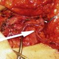
The surgeon begins by incising the edge of the skin flap. Initially, only one skin edge is incised to permit alteration of the skin paddle according to the feeding vessel selected. When mobilizing the flap and approaching the main pedicle, several additional subcutaneous vessels of more than 0.5 mm in diameter should be preserved ( Fig. 1 ). These additional vessels can be dissected to a reasonable length for anastomosis (often in the order of 1–4 cm) in case they are required at a later stage to augment arterial or venous flow. The flap can be beveled as necessary to provide additional tissue, with minimization of the skin paddle and donor site defect.
The dissection proceeds at either the suprafascial or subfascial level, depending on the flap being raised and surgeon preference. Small vessels can be identified within the flap as the dissection proceeds and these can be followed until they converge on the vessel of origin. The surgeon can use loupe magnification to assist in identification of vessels. Several intraoperative factors indicate the caliber of the perforator encountered. Factors include the size of the converging branches, whether the perforators have any visible pulsation, and also the extent of the facial opening traversed by the perforator. Larger fascial openings tend to be associated with larger perforators, even if this is not immediately apparent because of vessel spasm. It is crucial to approach the vessels in a blood-free environment because this assists in identifying the converging prefascial branches. Furthermore, the relative transparency of the subcutaneous fat allows the approximate location of the perforator to be viewed in advance ( Fig. 2 ), thus avoiding inadvertent damage. Blood-free clean dissection can be achieved by separating the tissue with low-current electrocautery and a wide spatula tip. It is essential to preserve all perforating vessels until a more dominant vessel is encountered. This precaution avoids the inadvertent transection of a dominant perforator that would compromise perfusion. If more than one perforator is to be included in the flap, then the perforators can be approached from different directions to allow simultaneous dissection and identification. The inclusion of multiple perforators must not sacrifice muscle or divide important motor nerves, which would negate the advantages of raising a perforator flap. If it is essential to divide a motor nerve then it must be surgically repaired. It is at this stage in flap dissection that accurate preoperative imaging allows speedier vessel dissection and sacrifice of nondominant vessels before reaching the predetermined dominant perforator.
Dealing with the deep fascia
Once the perforator has been identified and approached in the prefascial plane, further undermining should be performed between the subcutaneous fat and deep fascia for a distance of 2 to 4 cm around the perforator ( Fig. 3 ). It is crucial to perform this step before opening the deep fascia because once the fascia has been breached, the dissection becomes increasingly difficult to execute. In addition, the step provides an easily visible safety zone around the perforator, thus helping to avoid potential damage to the vessel when the remaining flap is lifted off the deep fascia in the final phase of dissection.
Once a clear circumferential view of the perforator has been established, it is necessary to open the deep fascia and follow the vessel through its intramuscular course. Opening the fascia is considerably easier if the fascial opening is large. The collagenous cuff around the perforator is cut and opened with specially designed fine scissors (Blondeel scissors, S&T AG, Neuhausen, Switzerland) until the loose connective tissue around the perforator is encountered ( Fig. 4 ). While moving and contracting, the loose connective tissue allows the perforator to glide between the muscle fibers and tendinous inscriptions of the rectus abdominis muscle. Dissection close to the vessels and within this loose connective tissue facilitates liberation of the vessels in a bloodless environment. Small fascial openings are often closely adherent to the perforator. In this situation, it may be necessary to incise the deep fascia lateral to the perforator and identify the immediate intramuscular course and then leave a cuff of fascia around the vessel. When incising the fascia, it is important to realize that the perforator may run obliquely under the fascia before diving into the muscle. Hence, diligence is required to avoid inadvertent damage to the vessel.
In certain flaps, the perforators can be approached from underneath the deep fascia, such as in the SGAP flap. In such situations, a safe and slow approach to the perforator is hampered by the presence of thick epimysia that need to be cut throughout their entire length. By selecting a subfascial approach, the perforator can be easily identified because of the transparent epimysia.
After identifying the main perforator below the deep fascia and just before entering the muscle or septum, the same circumferential dissection around the perforator should be done as above the fascia ( Fig. 5 ). This space is avascular and filled with loose collagenous tissue, so dissection advances easily. In this case, it is also important to release the remaining attachments of the deep fascia to the perivascular tissue cuff, freeing the perforator entirely above the muscle.
The intramuscular dissection
Once the initial incision of the fascia is made cranial and caudal to the perforator, the fascia can be opened to achieve better exposure. This step can be done in any direction but is preferentially performed in the same direction as the underlying muscle fibers and toward, or over, the main supplying deep vessels (see Fig. 5 ). It is crucial to incise the deep fascia as much as necessary to gain wide exposure of the vessels in the surgical field. The opening of the fascia leaves no donor site morbidity (provided proper resuturing is performed), whereas an inadequate exposure risks damaging of the vessel. At the point where the perforator enters the muscle, the muscle fibers are split in both directions by blunt dissection ( Fig. 6 ). The loose connective tissue cuff around the vessels guides the path through the muscle and indicates where the muscle fibers need to be split. In a first phase, the muscle fibers on top of the perforator and the axial vessels are split to expose the course of the pedicle through the muscle. This so-called deroofing process allows quick visual inspection of the pedicle to confirm its patency and valid vessel size, allows for early and quick adjustments to the intraoperative surgical strategy, but most of all allows wide exposure and thus good visualization of the entire pedicle ( Fig. 7 ).
Intramuscular dissection requires a meticulous technique to identify the main vessels and ligate any side branches. Accidental rupture, damage, or division of these branches results in bleeding, which obscures the operative view and risks damage to the vessels while achieving hemostasis. It is therefore essential to maintain a bloodless field at all times. Hemostasis should be secured with bipolar cautery or by clipping larger vessels, while constantly irrigating with warm saline to ensure that there is an adequate view of the vessel. Every side branch should be coagulated, ligated, or clipped at least 1 to 2 mm away from the perforator to avoid damage to the vessel and allow hemostasis to be resecured if there is continued bleeding.
Adequate exposure is also essential and is achieved with the use of self-retaining retractors or elastic retractor hooks (5 mm Sharp Hook Elastic Stays; Lone Star Medical Products, Houston, TX, USA). These retractors consist of a metal hook attached to a length of elastic. The hook retracts the tissue while tension is maintained by securing the elastic to a drape or skin staple. Retractors allow surgical instrumentation within the operative field to be kept to a minimum and free the surgical assistant for performing diathermy and irrigation. The perforator is always kept under direct vision to ensure that there is no tension on the vessel during flap manipulations and that it does not become desiccated. Securing the flap to the patient with staples or sutures should help prevent such inadvertent damage.
Direct handling of the vessels should be avoided, and while the perforators are completely freed from the surrounding tissue, the direction of dissection should always be sweeping away from the vessel itself. The dissection plane is at the level of the loose connective tissue layer that surrounds the vessels. This layer can be easily freed by blunt dissection, and any resistance encountered indicates a side branch. The instruments used for dissection are a matter of surgeon preference, but it is the authors’ practice to use bipolar diathermy forceps and dissection scissors, such as the Blondeel dissection scissors. The ring handle configuration of these scissors makes them suitable for preparation of fine structures, while the additional round handle and spring instrument configuration of the curved blades is extremely useful for fine trimming and dissection work. Once the larger deeper vessel is reached, dissection proceeds until sufficient caliber and length are achieved ( Fig. 8 ).
The main pedicle
Nerves will often be encountered at the level of the deeper vessels, often just underneath or within the deeper part of the muscle (see Fig. 6 ). Depending on the region of dissection, nerves can be classified as sensory, motor, or both. Adequate anatomic knowledge and nerve stimulators can help differentiate between these different fibers. Sensory nerves to the flap can be used to reanastomose to recipient sensory nerves. The length of the donor nerve can be increased as necessary by retrograde dissection. Motor nerves are dissected off the vessels over an adequate length to allow easy dissection of the vessels underneath. These nerves are always accompanied with one or more vascular side branches that need to be clipped. Occasionally, it is necessary to divide the motor nerve. In such circumstances, a quick reanastomosis can be performed with two 9-0 nylon stitches in the perineurium. This procedure is performed once the flap has been harvested or moved.
In a free perforator flap, dissection of the main vessel continues until an adequate pedicle length has been achieved to perform easy anastomosis. It is equally important to reach a donor vessel diameter that is suitable in size for the recipient vessels. Good interaction between the 2 teams harvesting the flap and preparing the recipient site saves valuable operative time and permits simultaneous identification and characterization of the recipient vessels. Often 1 artery and 2 comitant veins are included in the pedicle. The smallest comitant vein is immediately ligated at the end of the dissection of the pedicle to redirect and precondition flow into the larger vein. Transection of the vein is preferentially done immediately downstream of a branch interconnecting both comitant veins, the so-called H-connectors. Although multiple H-connectors exist over the entire pedicle, anastomosis at this site allows easy flow over the anastomosis. A good anastomosis of 1 vein is preferred over linking 2 veins to ensure appropriate venous flow.
Pedicled perforator flaps are planned as rotation, advancement, or transpositional flaps. The flaps may include one or more perforating vessels. However, flaps that include only one perforator offer increased freedom of movement without vascular compromise.
Once the feeding vessel has been identified, dissection of the vessel is continued as long as necessary to allow tension-free positioning of the flap in the recipient site’s defect. Tracing the perforator to its original source vessel is not always necessary once sufficient flap mobility is obtained. The aim is that when the flap is in the recipient site, there will be a degree of redundancy in the vessels, which ensures that there is no tension on the perforator, even when subjected to postoperative swelling or hematoma.
Rotation flap
In this situation, the dominant perforator acts as a pivot point. The flap is designed in the fashion of a propeller blade. The longest part of the flap turns approximately180° into the defect. Dissection of the perforator is often over a short distance but long enough to ensure that turning of the flap does not cause torsion of the vessel to the extent that it compromises perfusion. To sufficiently liberate the pedicle, the distance between the end of the dissection and the entry point in the flap needs to be slightly shortened. In that way, the vein is allowed to gently turn around the artery.
Advancement flap
In this case, the flap is simply slid in one direction, parallel to the direction of the muscle fibers. Dissection can go down to the axial vessels, which occasionally also require mobilization. An oblique course of the vessels to the skin surface is needed to allow movement of the flap. The longer the perforator the more the movement of the flap. Any design of skin island can be used, provided the limits of viability are respected.
Transposition flap
For further displacements, a longer leash is necessary. Movements of displacement, advancement, and rotation are performed simultaneously. Sufficient slack of the pedicle is necessary without causing kinking of the pedicle. It is essential to plan accurately and take exact measurements before making skin incisions. The size of the skin island is, once again, determined by the limit of blood flow to the periphery of the flap. Therefore, a central location of the perforator in the flap is preferred. It is also important to avoid subcutaneous tunneling of flaps in areas of dense subcutaneous tissues, such as the sacral and gluteal area. Subcutaneous tunneling may compromise perfusion, resulting in fat necrosis or in the worst instance, flap loss.
Recipient Vessel Selection and Preparation
Characteristics of the ideal recipient vessels include nontraumatized, nonscarred, nonirradiated, and disease free (at least 1 artery and 1 vein) with a sufficient length to perform easy microanastomosis and a diameter that corresponds to the donor vessels. Traumatized or irradiated vessels are difficult to expose, resulting in considerable risk of iatrogenic trauma and higher rates of thrombosis and flap failure. For some conventional free flaps, an interposition graft may be required to lengthen the flap pedicle and allow anastomosis out with the zone of injury. Many studies have shown the use of such grafts increases the rate of flap failure.
Unlike conventional free flaps, perforator flaps have consistently longer vascular pedicles that give them the increased flexibility for anastomosis to most recipient vessels. Thus, using a perforator flap can ensure that the most appropriate recipient vessels are selected to maximize the chance of flap success. The only limitation is the size of the flap vessels, which may necessitate an end-to-side anastomosis—the use of perforator recipient vessels or anastomosis to the side branch of a large recipient vessel. In supermicrosurgery, a perforator-to-perforator anastomosis that eliminates the need to dissect the flap vessels down to the larger axial vessels is performed. Such an anastomosis not only increases the ease of dissection but also makes the size of vessels more compatible. Although this may seem like an obvious progression, it should be remembered that usage of even smaller vessel diameters increases the risk of thrombosis and flap failure.
Preparation of the recipient vessels requires the same meticulous technique as flap raising. Hemostasis must be controlled by vascular clips or bipolar coagulation and aided by irrigation. Perivascular hematoma has been shown to cause vasospasm and flow disturbances, as well as prolonging vessel wall ischemia with resultant increased inflammatory response.
Exposure of the vessel should be wide to facilitate dissection and prevent compression of the pedicle against adjacent structures. Most part of the vessel dissection is performed under loupe magnification, with the final stages sometimes requiring an operating microscope, especially in a case of perforator-to-perforator anastomosis. The key to vessel dissection is advancement in the perivascular plane between the vessel and its vascular sheath. Once the sheath is divided, it can be retracted by gentle pulling and is then removed. The vessel is then sharply divided and the adventitia is trimmed. It is important to facilitate the anastomosis as much as possible by preparing the field adequately. A green or blue background is placed behind the vessel to be anastomosed, keeping all other structures out of the way. Continuous low–negative-pressure microsuction can be secured under the background using a small drain held with staples. Skin retraction is achieved with the autoretractors mentioned earlier. The flap is secured atraumatically in a suitable position by wrapping it in a saline-soaked gauze and stapling the gauze to the skin around the recipient site. All these maneuvers free the assistant to help with vessel preparation. Irrigation is performed with normal saline or heparin solution (50,000 U in 500 mL lactated Ringer solution) as long as the intima is exposed. Either the artery or the vein can be repaired first, and the sequence is often determined by vessel position rather than an optimal order. Vessels located in deeper planes or furthest away from the surgeon’s hands are sutured first. Two venous anastomoses may seem to be an obvious option if there is any doubt about venous outflow. However, caution should be exercised as this potentially reduces the venous outflow in each vessel and can lead to sludging and thrombosis.
Related posts:


Stay updated, free articles. Join our Telegram channel

Full access? Get Clinical Tree


