Fig. 23.1
Syringoid eccrine carcinoma. An indurated withish plaque on the cheek
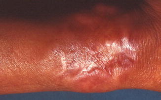
Fig. 23.2
Syringoid eccrine carcinoma. An infiltrated plaque with ill-defined margins on the wrist
Pathology
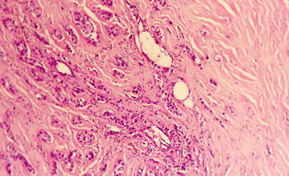
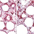
The histological appearance includes an infiltrative growth pattern with formation of tubules, small elongated nests and cords, syringoma-like tadpole structures embedded in a desmoplastic stroma (Figs. 23.3, 23.4 and 23.5). These structures are lined with a double layer of cells with flattened cuboidal or squamous differentiation. There is prominent squamoid differentiation, most apparent superficially, where neoplastic aggregates are larger and composed of epithelial cells with abundant amphophilic cytoplasm. Cellular atypia and mitoses are generally mild to moderate. Perineural and intraneural invasion is a characteristic finding. This tumor extends deep into the dermis, subcutis, and skeletal muscle and sometimes into the bone. The tumor cells express typically cytokeratin AE1/AE3, CAM 5–2, Leu M1, CEA (Fig. 23.6), and PS100.
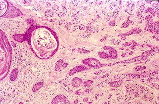
Fig. 23.3
An infiltrative proliferation composed of small elongated nests and cords, squamoid islands with keratinous cyst and syringoma-like tadpole structures with clear cell changes embedded in a desmoplastic stroma