Fig. 27.1
Primary mucinous carcinoma of the skin. A dermal tumor consisting of small islands of epithelial tumor cells “floating” in large pools of slightly basophilic mucin separated by thin fibrovascular septa (Courtesy of D. Kazakov, Pilsen, Czech Republic)
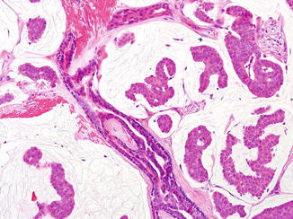
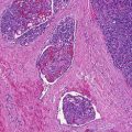
Fig. 27.2
Primary mucinous carcinoma of the skin. The clustered epithelial cells are small and uniform with cuboidal nuclei and can form ductal structures in the epithelial islands floating in large pool of mucin (Courtesy of D. Kazakov, Pilsen, Czech Republic)
Differential Diagnosis
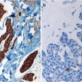
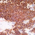
The clinical differential diagnosis is broad and includes other adnexal tumors, basal cell carcinoma, squamous cell carcinoma, sebaceous carcinoma, and epidermoid cyst. Histological differential diagnosis includes metastatic adenocarcinomas, particularly of the breast and gastrointestinal tract, mucinous basal cell carcinomas, and other sweat gland carcinomas. The mucin in other sweat gland carcinomas is usually a sulfated mucopolysaccharide with different staining properties. Metastatic mucinous adenocarcinoma may be histologically indistinguishable from PMCS. A few subtle histological features should be searched for and may provide help in establishing the correct diagnosis: mucinous carcinomas have more mucin, more clusters of epithelial cells, fewer ductal structures, fewer solid areas, and fewer mitoses than metastatic tumors. An additional clue for a primary lesion is the presence of an in situ component identified as epithelial islands being bounded by a myoepithelial layer, which is highlighted by p63, CK 5/6, calponin, SMA, and HHF-35. Immunohistochemical staining and cytokeratin profiles are also of great help in the differential diagnosis. PMCS shows positive cytoplasmic staining with S-100, carcinoembryonic antigen (CEA), vimentin, epithelial membrane antigen (EMA), α-actin, and cytokeratin 7 (CK7), as well as with estrogen receptor and progesterone receptor. CK20 is useful to differentiate metastatic lesions from the intestinal tract since it is negative in PMCS. In addition, “dirty necrosis” is frequently found in intestinal mucinous adenocarcinomas involving the skin. In mammary mucinous adenocarcinoma involving the skin, lesions are on the chest wall, breast, and axilla, and these locations can serve as clue to the breast origin.
Related posts:


Stay updated, free articles. Join our Telegram channel

Full access? Get Clinical Tree


