Fig. 57.1
Primary cutaneous ES/PNET displays a nodular dermal-based proliferation of small round blue cells (Image courtesy of Steven D. Billings, MD)
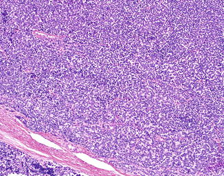
Fig. 57.2
ES/PNET is composed of hypercellular sheets of monotonous oval/round cells
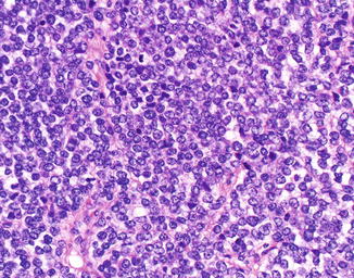
Fig. 57.3
The tumor cells have round to oval nuclei, vesicular chromatin, and minimal cytoplasm. Multiple mitoses are present
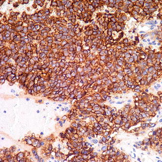
Fig. 57.4
Strong diffuse membranous expression of CD99 is characteristic of ES/PNET
Differential Diagnosis
The most important differential diagnoses to exclude are cutaneous metastasis of ES/PNET and cutaneous extension of a deep soft tissue ES/PNET; both require clinical and radiologic correlation. Additionally, the histological differential diagnosis for primary cutaneous ES/PNET is quite broad and includes other “small round blue cell” tumors that may be present in the skin and subcutis such as neuroblastoma, embryonal or alveolar rhabdomyosarcoma, lymphoblastic lymphoma, leukemia cutis, carcinoma (poorly differentiated adnexal carcinoma including malignant trichoblastic neoplasms, Merkel cell carcinoma, metastatic small cell carcinoma, other neuroendocrine carcinomas), small cell melanoma, desmoplastic small round cell tumor, or poorly differentiated synovial sarcoma. A combination of immunohistochemistry and molecular analysis can usually exclude these entities and confirm the diagnosis of ES/PNET. It is worth noting that while strong diffuse membranous expression of CD99 is a highly sensitive marker of ES/PNET, it is not entirely specific; CD99 expression may be seen in many other small round blue cell tumors. Additionally, while RT-PCR may specifically confirm the presence of an ES/PNET-specific gene rearrangement, FISH showing EWS gene rearrangement is not entirely specific for ES/PNET, as the EWS gene may be rearranged in a variety of other neoplasms including myoepithelioma, myoepithelial carcinoma, desmoplastic small round cell tumor, clear cell sarcoma, and angiomatoid fibrous histiocytoma among others. Correlation with clinical and histological features is essential when interpreting the results of molecular analysis.
Related posts:


Stay updated, free articles. Join our Telegram channel

Full access? Get Clinical Tree


