Fig. 47.1
Signet ring cell melanoma. Low-power magnification demonstrates an intraepidermal proliferation of atypical melanocytes with pagetoid spread. Within the dermis, there are sheets of atypical melanocytes, some of which demonstrate signet-ring morphology
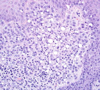
Fig. 47.2
Signet ring cell melanoma. High magnification delineates enlarged pleomorphic cells, some of which have a single intracytoplasmic vacuole. The nucleus is pushed eccentrically and is indented. A dermal mitotic figure is evident. These cells expressed MART-1
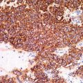
Immunohistochemical staining may be necessary to diagnose melanoma with signet-ring cells. Typically, the tumors express S-100 protein, although rarely, tumors lacking S100 protein expression or only demonstrating focal positivity are reported. Other conventional melanocytic markers such as HMB45 and Mart-1 may be expressed but are less frequently positive.
Differential Diagnosis
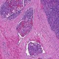
Signet-ring cells may be encountered in benign melanocytic nevi as well as melanoma (Fig. 47.3), and as such, the presence of such morphology should not automatically trigger a diagnosis of malignancy. Signet-ring cell melanoma also has overlapping histological features with other histological variants of melanoma, including rhabdoid and balloon cell melanoma. In melanoma with rhabdoid features, the nucleus is pushed to the side, but the cytoplasm acquires a dense pink, eosinophilic rather than clear appearance, thereby mimicking primitive rhabdomyoblasts. In contrast, balloon cell change, like signet-ring change, is characterized by clear cytoplasm within the melanocyte, but the nucleus is central, rather than eccentrically placed, and there may be multiple intracytoplasmic vacuoles. Balloon cell change is thought to represent degeneration and overproduction of melanosomes, and electron microscopy does not demonstrate accumulation of vimentin filaments.
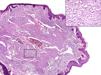
Fig. 47.3
Signet-ring cell change in a dermal nevus. Scanning magnification shows a predominantly intradermal nevus. Focally (inset), signet-ring cells are present
Signet-ring cells are classically described in mucin-secreting adenocarcinomas, and melanoma with a predominant signet-ring cell pattern can closely mimic such a tumor. The presence of intracytoplasmic mucin or expression of cytokeratin would support a diagnosis of signet-ring adenocarcinoma, whereas the absence of mucin or cytokeratin expression or the presence of associated pigment would favor a melanoma. Immature adipocytes (lipoblasts) may sometimes mimic signet-ring cells and can be distinguished by lipid material within the cytoplasm and nuclear vacuolization. Signet-ring cells have also been described in vascular tumors such as hemangioendothelioma, some epithelioid smooth muscle tumors, and lymphomas. In such cases, immunohistochemical stains may be necessary to determine the tumor cell derivation. Other primary cutaneous neoplasms, including squamous cell carcinoma, basal cell carcinoma, and adnexal tumors, can sometimes exhibit signet-ring cells.
Related posts:


Stay updated, free articles. Join our Telegram channel

Full access? Get Clinical Tree


