Fig. 67.1
Subcutaneous GD-TCL: Infiltrated and ulcerated plaque on the arm (By courtesy of Dr. B. Pfeiff, Germany)
Pathology
CGD-TCL shows epidermotropic, dermal (diffuse or nodular), and/or subcutaneous infiltrates. The subcutaneous form displays predominantly lobular infiltrates (Fig. 67.2). The neoplastic cells are of variable size, mostly medium-sized to large with irregular chromatin-dense or vesicular nuclei (Fig. 67.3). Angioinvasive growth and necrosis are common features. CGD-TCL displays a CD2+ CD3+ CD56+ phenotype with expression of cytotoxic molecules (TIA-1, granzyme B, perforin) (Fig. 67.4). The tumor cells are often CD4/CD8 double negative. By definition, the neoplastic cells express TCR γ/δ and lack TCR α/β (βF1). The γ/δ+ TCR phenotype can be demonstrated by expression of TCR delta-1 (TCRδ+) on fresh-frozen tissue or TCR gamma (TCRγ+) on formalin-fixed, paraffin-embedded sections. Molecular studies reveal clonal rearrangement of TCR gamma or delta genes. The presence of isochromosome 7q is a common genetic abnormality. EBV is generally negative.
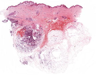
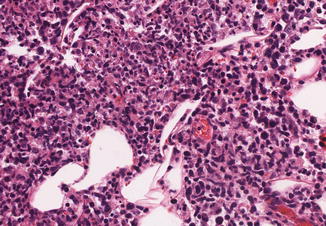
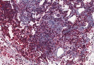
Fig. 67.2
Subcutaneous GD-TCL: Predominantly subcutaneous lobular infiltrates
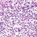
Fig. 67.3
Subcutaneous GD-TCL: Infiltrates of small- to medium-sized lymphocytes with atypical chromatin-dense nuclei
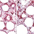
Fig. 67.4
Subcutaneous GD-TCL: Expression of CD56 by tumor cells
Differential Diagnosis
Histologically epidermotropic forms of CGD-TCL simulate mycosis fungoides (MF) and its subtype pagetoid reticulosis (PR), which may occasionally express a γ/δ+ TCR phenotype, but otherwise show classic clinical and histological features of MF (non-erosive patches and plaques) and PR (solitary plaque), respectively, and have an indolent course. The subcutaneous form of GD-TCL has to be distinguished from subcutaneous panniculitis-like T-cell lymphoma, which is defined by the expression of a TCR α/β+ phenotype as the main discriminating feature. The latter shows a favorable prognosis which contrasts to the aggressive course of GD-TCL. Further differential diagnosis of subcutaneous GD-TCL includes extranodal NK/T-cell lymphoma, nasal type, which is associated with EBV and displays a CD3ε+ CD56+ phenotype.
Related posts:


Stay updated, free articles. Join our Telegram channel

Full access? Get Clinical Tree


