Fig. 41.1
Myxoid liposarcoma. A large, slowly growing, deeply located painless mass on the upper back
Pathology
Myxoid LPSs are well circumscribed or encapsulated, lobulated soft tissue tumors (Fig. 41.2). They are composed of a proliferation of small, bland, stellate, spindle-shaped or round cells, with small vacuoles dispersed in a myxoid matrix, giving a pseudolymphangiomatous appearance. A complex plexiform arrangement of small thin-walled capillaries with a chicken wire or crow’s feet distribution represents a characteristic feature of myxoid LPS (Fig. 41.3). Multivacuolated lipoblasts are easily identified in the more cellular peripheral regions (Fig. 41.4). Mitoses may be found but are usually sparse. Pleomorphic and multinucleated cells may occasionally be seen. Granular eosinophilic hibernoma-like cells and leiomyomatous, cartilaginous, and osseous metaplasia may occasionally be encountered. Myxoid LPS may present with more cellular areas composed of a variable number of oval-to-round larger cells with hyperchromatic nuclei and inconspicuous cytoplasm (Fig. 41.5). When the round cell population is less than 10 %, the tumor should be regarded as a low-grade liposarcoma. In contrast, when the round cell component predominates, the tumor is associated with a more aggressive behavior (Fig. 41.6). This tumor is known as either combined and round cell LPS or high-grade myxoid LPS. Hemorrhage and/or necrosis may be present in association with high-grade histological features and portend a poor prognosis.
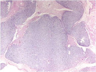
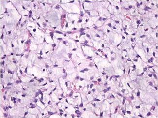
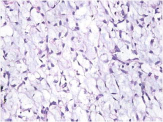
Fig. 41.2
The tumor is composed of well-circumscribed lobulated masses
Fig. 41.3
A chicken wire or crow’s feet distribution of vessels is a characteristic feature
Fig. 41.4


The presence of multivacuolated lipoblasts is an important diagnostic feature
Related posts:


Stay updated, free articles. Join our Telegram channel
Full access? Get Clinical Tree
