Reference
Number of patients
Type I
Type II
Type III
Number (percent)
Number (percent)
Number (percent)
Upton [29]
68
28 (41 %)
24 (35 %)
16 (24 %)
Cohen and Kreiborg [31]
44
20 (45 %)
18 (39 %)
6 (16 %)
Holten et al. [32]
45
29 (64 %)
10 (22 %)
6 (13 %)
Chang et al. [33]
10
5 (50 %)
1 (10 %)
4 (40 %)
Fearon [34]
17
11 (65 %)
2 (12 %)
4 (24 %)
Guero [35]
52
11 (21 %)
19 (37 %)
22 (42 %)
Totals
236
104 (44 %)
74 (31 %)
58 (25 %)
Treatment
Reconstruction of the hand in patients with Apert syndrome is an evolving technique that presents a significant challenge to hand surgeons, and the treatment of the numerous hand anomalies encountered in Apert syndrome requires a complex operative plan with multiple stages through childhood and into adolescence. There has been a lively discussion in the literature over the past 20 years, adding to the prior body of literature, in which a variety of reconstructive plans have been outlined and modified. Although there are several common goals of each of these reconstructive plans, each author or group has their own preferences and biases. Several factors account for the lack of a clear consensus on the management of these patients, including the rarity of this syndrome, the presentation of each patient with a unique cluster of anomalies with varying degrees of severity, the role of surgeon preference and surgeon comfort in determining a reconstructive plan, and the difficulty in having the long-term follow-up needed to evaluate the durability of the reconstruction. Despite this lack of consensus, the common goals between most of the proposed reconstructive plans include minimizing the number of procedures, maximizing the functional outcome of the hand, and providing a favorable cosmetic result, which includes preserving as many digits as possible through judicious use of amputations.
It should be noted that in the past, there was some question about the utility of offering hand reconstruction to Apert syndrome patients due to mental impairment that can be quite severe. However, we feel and want to echo the sentiment of other authors [8, 35] who also specifically have emphasized the point that, regardless of the degree of mental impairment of the patient, the functional gains and cosmetic improvements following reconstruction offer significant quality of life improvements, both for the patient and for the family, that should not be withheld from Apert syndrome patients.
The technical goals for reconstruction of the Apert hand address syndactyly and symphalangism, thumb radial clinodactyly, and later secondary deformities requiring revision. These goals have been organized by several authors into a reconstructive plan. Considerations that must be made in the formulation of a reconstructive plan include age of the patient at the time of the initial operation, timing and sequence of the release of border digits, creating skin flaps and providing soft tissue coverage, need for digital amputation, thumb lengthening and straightening, and secondary revisions.
Patient Age
Ideally, patients with Apert syndrome should be referred shortly after birth to a center with the multidisciplinary expertise necessary to treat the hand and craniofacial anomalies associated with Apert syndrome. However, due to a variety of reasons, including patients who were born in parts of the world without the multidisciplinary teams available for reconstruction, Apert syndrome patients are often seen well after infancy. This can present a challenge and requires modifications to the reconstructive sequence in these patients.
The age of the patient is particularly relevant to the decision of whether both hands are operated on simultaneously or whether the same operation for each hand is delayed in a staged manner. Following each reconstruction, the patients are typically placed in casts or splint, which is variable from group to group. In patients who require bilateral upper extremity restraints, this can cause significant distress for the patient, depending how independent and interactive he or she is, and place a significant burden on the parents, again, depending on how dependent the patient is on the parents for assistance with basic tasks of daily care. The age below which operations are performed on bilateral extremities simultaneously varies from 12 [33, 35, 36] to 18 [37] to 24 months [8] among authors who specified. In patients who underwent the same procedure on each hand individually, the delay between procedures on each hand ranged from as short as 2 weeks [35] up to 3 [33] to 6 months [35, 36] to allow time for the contralateral hand to heal and become more functional.
Another consideration for timing, though mentioned in only one paper, was discussed by Hoover et al., who recommended waiting 6–9 months before operating on the same hand to allow time for adequate revascularization [8]. However, many authors do not wait this long and have not reported increased complications due to vascular compromise.
Syndactyly, Symphalangism, and Border Digits
Timing of release of the border digits is a source of controversy. Some authors suggest that postponing separation of the digits will lead to angular growth deformities due to differential growth of each of the digits [8, 29, 38], while others state that in their experience this is not the case [34]. Another consideration in the timing of the release of the digits is to provide early mobility to promote earlier motor development. Earlier release of the thumb and the small finger, the border digits, allows the patient to begin development of a grasp. Hoover recommends performing a border digit release by 1 year of age [8]. Fearon, however, did not observe these problems in his patients that did not undergo early border digit release [34].
For those authors that prioritize the release of the border digits in Upton Type II and III hands, two additional procedures are required to release the remaining syndactylies. This is the case because the remaining syndactylies after release of the border digits are the index-long and long-ring finger syndactylies. Releasing both of these syndactylies in the second and third web spaces requires operating on both sides of the long finger. Operating on both sides of the long finger during the same operation theoretically risks compromising the vascular supply to the long finger and having a shortage of flap skin [30, 34, 36]. To minimize this risk, the long finger syndactyly release is typically staged as two separate operations, which increases to three the number of operations a patient must undergo and increases the time spent by a patient without full release of all of his or her fingers. To reduce the number of operations, most surgeons release alternating web spaces, including releasing one side of the long finger syndactyly during the first operation while neglecting one of the two border digit syndactylies during the first operation [34].
As just mentioned, the concern for vascular compromise dictates operative staging and forces surgeons to choose either prioritizing border digit release or limiting the number of operations to two. Even with careful consideration of the vascular supply to the digits, the aberrant anatomy of the neurovascular bundles increases the risk of inadvertent disruption of the blood supply to the digits. To address these problems, Harvey et al. examined the role of CT angiogram to assist with mapping of the vascular supply to each digit [39]. This imaging was done concurrently with CT imaging performed for operative planning for craniofacial reconstruction. After mapping the vascular supply to the hand and planning the surgical approach, they attempted to perform a single-stage syndactyly release paying careful attention to the vascular anatomy based on the CT angiogram findings. In both hands of all five patients in this study, they were able to perform successfully a single-stage syndactyly release without any major complications.
We have not adopted this approach because another problem with release of adjacent fingers is the shortage of dorsal skin that can be used for dorsal flap coverage of the webs. Therefore, we feel that the risks and limitations of adjacent finger release outweigh the benefit of a single-stage approach.
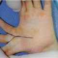
Separation of the syndactyly in the fingers is typically performed with a zigzag incision. This results in interdigitating triangular flaps along the sides of the newly released digits. The purpose of this pattern is to avoid a straight-line scar along the sides of the fingers due to the concern for scar contracture leading to deviation of the finger or limitation of function. Syndactyly release in Apert syndrome is different because the fingers have some degree of symphalangism, with resultant stiff joints that will not deviate with scarring of the skin incisions [34]. Straight-line syndactyly release incisions will prevent the zigzag incisions from extending onto the dorsal and volar surface of the fingers and will allow application of one piece of skin graft to each side of the finger (Fig. 14.1) Upton suggests the small finger should be treated with extra caution with regard to the use of straight-line incisions.
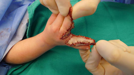
Fig. 14.1
Full thickness skin grafting after straight-line syndactyly release
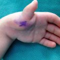
Because many syndactylized fingers in Apert syndrome are complex (involving bone at the tip), two specific operative maneuvers are critical. Zigzag fingertip flaps, attributed to Buck-Gramcko, are useful for recreating the nail folds [40] (Fig. 14.2). Also, intraoperative fluoroscopy is used to visualize the bony fusion prior to osteotomy. A fine gauge needle is placed slightly off center to the proposed longitudinal osteotomy, and the osteotome is slid on top of the needle to allow precise sectioning of the bone (Fig. 14.3).
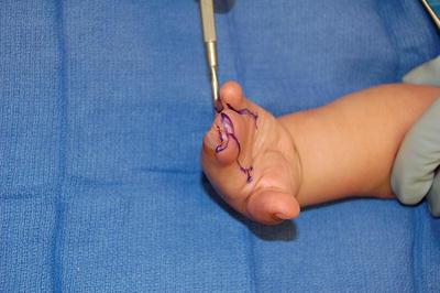
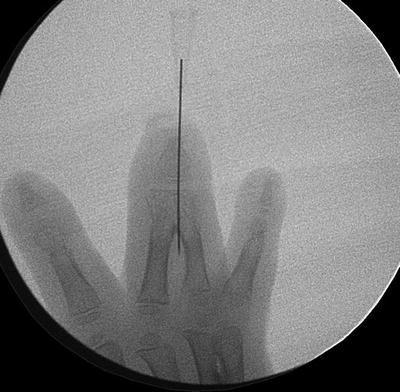
Fig. 14.2
Markings for zigzag fingertips for recreating the nail folds
Fig. 14.3
Fluoroscopy image demonstrated needle positioning used to guide longitudinal osteotomy
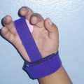
Several flaps have been described for reconstruction of the second, third, and fourth, web spaces. Barot and Caplan describe a dorsal rectangular flap that they inset into a volar T-incision [36]. Guero describes an omega-shaped dorsal flap [35]. Other authors perform a similar long dorsal flap for reconstruction of the web space. Fearon, however, uses equal length triangular dorsal and volar flaps [34]. This results in a length-to-width ratio that provides more favorable blood supply to the distal tip of the flap and better healing. He attributes this technique as the reason for his very low reported rate of 3 % for secondary syndactylies requiring reoperation. He designs the base of his dorsal flap proximal to the base of the volar flap to recreate the normal slope of the web space. In Upton’s commentary on Fearon’s article, Upton agrees with the Fearon’s triangular flaps, but he cautions that the second web space may require a future secondary release due to increased metacarpal growth [34]. To accommodate for this, Upton recommends considering a wide rectangular flap being used initially, which can then more easily be advanced again if needed later in life. This is the flap design that we usually choose to use (Fig. 14.4).
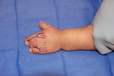
Fig. 14.4
Rectangular dorsal advancement flaps for web space reconstruction
For areas along the fingers that are not covered by the skin flaps raised during release of the syndactyly, full thickness skin grafts are typically applied. Split thickness skin grafts are rarely used due to graft contraction leading to decrease in size of the web space and due to the risk of recurrence of syndactyly. Full thickness skin grafts are typically harvested from the groin crease, avoiding the future hair-bearing skin, or occasionally from the antecubital crease. Skin harvested from circumcisions should never be used due to darkening of the harvesting skin with time, which provides a poor cosmetic result that patients often request to be revised. In cases with small areas of exposed bone without overlying vascularized tissue in the distal half of the released digits, Fearon did not provide coverage with skin grafts or tissue flaps [34]. This reduced the need for full thickness skin graft tissue but without increasing wound healing complications. In addition to skin grafts, several other techniques of increasing complexity have been suggested for providing soft tissue coverage, including pedicle groin flaps [38], tissue expanders, and silastic sheets [41]. Although these were not used in the more recent large series, the reconstructive surgeon should remain mindful of these techniques should additional soft tissue coverage be needed.
The techniques for the pedicle groin flap and tissue expansion are rather evident, but the use of silastic sheets warrants further discussion. This technique was described by Stefansson and Stilwell for use in cases in which extensive bone and soft tissue remains exposed after separation of complex syndactyly [41]. They developed this technique for cases in which they were concerned that the exposed bed of bone and cartilage would be poorly suited to a full thickness skin graft. After separating the digits, they interposed a 1-mm thick sheet of silastic along the exposed bone and cartilage and then closed the skin flaps in their original positions, leaving the silastic sheet in place. The silastic sheet was removed 1 month later, at which time they noticed a well-formed capsule covering the previously exposed bone. They then applied a full thickness skin graft from the groin, which survived. This is not usually necessary as small areas of exposed bone can be covered by skin grafts.
Related posts:

Stay updated, free articles. Join our Telegram channel

Full access? Get Clinical Tree


