Fig. 1.1
A Linea alba B Linea semilunaris C Arcuate line
1.1 Linea Semilunaris
The linea semilunares can be seen as a pair of linear impressions in the skin that correspond with the lateral most edges of the rectus abdominis. It is formed by the band of aponeuroses of the external oblique, the internal oblique, and transverse abdominal muscles [2].
1.2 Arcuate Line
The arcuate line is defined by the most inferior extension of the posterior rectus sheath, forming a crescent-shaped border. The arcuate line is generally located two fingerbreadths from the umbilicus to midway between the umbilicus and pubis. There are reports in the literature, however, that state the arcuate line is closer to 75 % of the distance between pubic crest to umbilicus or 1.8 cm superior to the anterior superior iliac spine [3]. It is not visible from the exterior [3].
1.3 Linea Alba
In the midline, a slight furrow extends from the xiphoid process above, to the pubic symphysis below, representing the linea alba in the abdominal wall. The fibres of the anterior and posterior sheaths of the rectus muscle interlace at the midline, forming the linea alba.
From the surgical anatomical perspective, the abdominal wall consists of various structural layers.
1.4 Layers of the Abdominal Wall
From the surgical anatomical perspective, the abdominal wall consists of various layers, namely, the skin, superficial fascia, fat, muscles, the transversalis fascia, and the parietal peritoneum.
1.4.1 Skin and Subcutaneous Tissue
This outmost layer consists of the skin, superficial fat layer, the fascia superficialis (Scarpa’s fascia), and the deep fat layer.
1.4.1.1 Skin
Epidermis
The epidermis is formed of five layers, and the epithelial cells transform itself into a keratin layer, which constantly peels off.
Dermis
The deeper structure called the dermis has two layers with no threshold between them. They are:
The papillary layer which is thinner and external
The reticular layer which is deeper and denser
The skin covering the anterior abdominal wall is thin, in comparison with that of the back, and is relatively mobile over the underlying structural layers except at the umbilical region, where it is fixed. The thickness of the abdominal skin seems to augment when approaching the midline and especially around the umbilicus. Natural elastic traction lines of the skin (also known as skin tension lines or Kraissl’s lines) of the anterior abdominal wall are disposed transversely. Above the level of the umbilicus, these tension lines extend mostly in a horizontal direction, while below this level, they continue along with a slight inferomedial oblique direction. Incisions made along, or parallel to, these lines tend to heal without much scarring, whereas incisions that cross these lines tend to result in wide or heaped-up scars [1].
1.4.1.2 Superficial Fat Layer (Superficial Adipose Tissue)
Superficial fat, just under the dermis, is formed of large fat lobes encased between fibrous septa in a honeycomb-like structure and presents nearly constant characteristics throughout. These septa (retinacula cutis superficialis or Camper`s fascia) appear well defined. They are mostly oriented perpendicular to the surface and are mechanically strong, anchoring the dermis to the deeper planes (Fig. 1.2).
Fig. 1.2
Retinacula cutis superficialis or Camper’s fascia
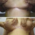
Stronger attachments to the dermis in the midline and above the umbilicus have been reported [4]. This is the reason why liposuction performed above the umbilicus and close to the midline is more prone to result in irregularities. The attachments of the retinacula cutis superficialis to the skin at 5–7 cm above the umbilicus can occasionally be stronger than usual and create a deep horizontal line. This deformity cannot be corrected by classical or scarpa-saving undermining – more superficial interventions are necessary (Fig. 1.3).
Fig. 1.3
The attachments of Camper’s fascia to the skin above the umbilicus can occasionally create a deep horizontal line
Things to Remember
In order to mimic this ideal anatomy and to create a beautiful result, the superficial landmarks are to be recreated.
Liposuction performed above the umbilicus and close to the midline is more prone to irregularities.
On rare occasions particularly above the umbilicus, the retinacula cutis superficialis’ attachments to the skin may be stronger than usual and create a deep horizontal line.
This deformity cannot be corrected by classical or scarpa-saving undermining – more superficial interventions are necessary.
1.4.1.3 Fascia Superficialis (Scarpa’s Fascia)
The superficial fascia comprises two distinct layers: an outer, adipose layer lying subjacent to the dermis and an inner fibroelastic layer termed Scarpa’s fascia, the membranous layer of superficial fascia [1]. The fibrous layer with a membranous appearance –the fascia superficialis – is continuous and well organized. It separates the superficial and deep adipose tissues (Fig. 1.4).
Fig. 1.4
Scarpa’s fascia
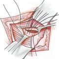
This layer can be followed as a dissection plane from the thorax to the inguinal ligament. It does not appear uniform in thickness. Being a well-defined white layer in the lower abdomen, thickening toward the inguinal ligament where a multilayered structure of multidirectional collagen bundles is perceptible. The Scarpa’s fascia loses consistency in the upper abdomen, where it can be identified as a much thinner translucid collagen layer through which adipose tissue can be seen [4]. This membrane, which is strongly fused medially to the linea alba and caudally to the inguinal ligament and the osseous prominence of the iliac crest, cranially continues into the thorax. The membranous layer (Scarpa’s fascia) is an important structure, which is strong enough to diminish the tension of sutures when identified and sutured in continuity during closure of the abdominal flap [5].
1.4.1.4 Deep Fat Layer (Deep Adipose Tissue, DAT)
Deep adipose tissue appears very different from the superficial adipose tissue, as its fat lobes are smaller, flatter, and less well defined (Fig. 1.5). This adipose layer shows significant variations in terms of thickness between different areas. Towards the points, at which the membranous layer of the subcutaneous tissue adheres to salient structures (e.g., the inguinal ligament, bony prominences, linea alba), they become thinner and tend to progressively reduce the fat component. However, the network of collagen fibres (retinacula cutis profunda) become stronger and more tightly packed and connects the deep aspect of the membranous layer to the deep fascia.
Fig. 1.5
Deep adipose tissue appears very different from the superficial adipose tissue, as its fat lobes are smaller, flatter, and less well defined
In the deep adipose layer, the fibrous septa are predominantly obliquely and horizontally oriented (retinacula cutis profunda) and connect the membranous layer (Scarpa’s fascia) to the fascia of the rectus abdominis or external oblique muscle [4]. The membranous layers DAT and SAT create a sliding system that absorb the mechanical stimulations applied to the skin or that are generated by muscular contractions.
In this way the subcutaneous tissue ensures autonomy between the skin and the muscles. If any scarring creates adhesion between the skin, membranous layer, and deep fascia, every muscular contraction could also affect the skin, activating the cutaneous receptors – also vice versa: every stimulation of the skin could be transmitted to the underlying structures. This may explain the importance of the correct layered reconstruction of the subcutaneous tissue in avoiding complications after closure of the abdominal surgery wounds [4].
Things to Remember
- 1.
A trilaminar structure is always present at the abdominal subcutis.
- 2.
Over the rectus abdominis muscle, there is a thicker region, and the difference is mainly attributable to the superficial compartment.
- 3.
The deep fat compartment has a minor contribution to the overall thickness, which is less than 25 % of the total thickness.
- 4.
The superficial fat compartment is more susceptible to increase in thickness in obesity compared with the deep compartment.
- 5.
The Scarpa’s fascia is always present and does not become vestigial with increased adiposity [6].
1.5 Musculofascial System of the Abdomen
The abdominal wall consists of five paired muscles: three flat muscles and two vertical muscles. The three flat muscles are the external oblique, internal oblique, and the transversus abdominis. The two vertical muscles are the rectus abdominis and pyramidalis. The three-layered structure, combined with extensive aponeuroses, works in a synkinetic fashion. Fusion of the fascial layers of these muscles forms three distinct fascial lines: the linea alba and two semilunar lines. The linea alba is formed by the fusion of both rectus sheaths at the midline, while the semilunar lines are formed by the union of the internal oblique, transversus abdominis, and external oblique as they join the rectus sheath (Fig. 1.6).
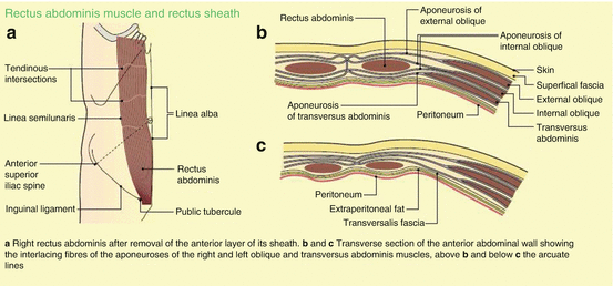
Fig. 1.6
Musculofascial system of the abdomen. (a) Right rectus abdominis after removal of the anterior layer of its sheath. (b, c) Transverse sections of the anterior abdominal wall showing the interlacing fibres of the aponeuroses of the right and left oblique and transversus abdominis muscles, above (b) and below (c) the arcuate lines (Source: Moore [19])
The abdominal muscular anatomy is well known with one vertical muscle anteriorly and three large lateral muscles overlying each other inversely. The vertical rectus muscle is divided by the linea alba.
1.5.1 Linea Alba
The linea alba is the rest of the embryonic ventral suture made up of three distinct aponeurotic layers originating from three lateral abdominal muscles, migrating to the midline, encircling the rectus abdominis muscle and fusing in the midline. It is a three-dimensional composition of tendon fibres from abdominal wall muscles. The cranial aspect is attached to the xiphoid process of the sternum, while caudally, it inserts at the pubic symphysis. This strong attachment to the sternum prevents any hyperextension of the vertebral structures. Also, the midline insertions of these fibres play a significant role in stabilizing the abdominal wall [2, 7].
Related posts:


Stay updated, free articles. Join our Telegram channel

Full access? Get Clinical Tree


