Seborrheic Keratoses
Silvia Soohyun Kim
Shang I. Brian Jiang
I. BACKGROUND
Seborrheic keratoses (SKs) are common benign epidermal tumors found in middle-aged and elderly populations. The term “seborrheic” refers to the lesion’s greasy appearance and location in areas that have many sebaceous glands. However, there is no known relationship to sebaceous gland function, seborrhea, or seborrheic dermatitis. The cause of SKs is unknown. Genetics (polygenic), sun exposure, and infection are all implicated as possible predispositions to developing SKs. In mature SKs, deoxyribonucleic acid (DNA) synthesis is decreased, while ribonucleic acid and protein synthesis is increased with irregularities in the expression patterns of apoptosis markers. Many patients with SKs have positive family history for the condition, but validated studies are lacking. Sun exposure has been shown to increase the prevalence of SKs, but there are some studies disagreeing with the role of genetics in SK development. Viral infection has also been explored as a possible cause of SKs. SKs from the genital region may contain human papillomavirus (HPV) DNA, but the role of HPV in developing SKs has been controversial. A recent study reported that HPV-positive genital SKs arise in younger, sexually active age groups.
While concern about these lesions is primarily cosmetic, some lesions with multiple dark colors raise the question of melanoma. Rarely, malignant lesions (i.e., basal cell carcinoma [BCC] and squamous cell carcinoma [SCC]) can arise within SKs, especially the reticulated type on sun-damaged skin. In a retrospective study of 813 histologically diagnosed SKs, 5.3% were associated with nonmelanoma skin cancer. No melanomas were observed. The most common malignancy was Bowen disease, followed by BCC, then invasive SCC.
II. CLINICAL PRESENTATION
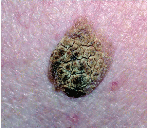
SKs are generally asymptomatic. Irritated lesions or those in intertriginous areas may cause intense pruritus. SKs start as small flesh-colored, yellow, or tan-colored waxy papules that may grow to become dark brown or black greasy verrucous lesions with a distinct border (Fig. 39-1). The rough scale may sometimes flake or be rubbed off but will regrow. The keratoses appear to be “stuck on” the skin (Fig. 39-2), and close inspection with a hand lens will expose the presence of horn cysts or dark keratin plugs. Stucco keratosis, a variant of SKs, is 1- to 5-mm lightly colored keratotic papules on the dorsa of the hands and feet and lower legs. A variant of SKs known as dermatosis papulosa nigra is seen primarily on the cheeks in blacks or other dark-skinned individuals with a familial predisposition. These lesions are small, pigmented papules that may be pedunculated.
 Figure 39-1. Seborrheic keratosis with “stuck-on” appearance. (From Goodheart HP. Goodheart’s Photoguide of Common Skin Disorders. 2nd ed. Philadelphia, PA: Lippincott Williams & Wilkins; 2003.) |
Stay updated, free articles. Join our Telegram channel

Full access? Get Clinical Tree


