Fig. 14.1
Sebaceous carcinoma. A slow-growing, painless pinkish nodule on lower tarsal plate
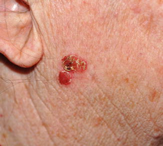
Fig. 14.2
Sebaceous carcinoma. An erythematous slowly enlarging nodular plaque with a yellowish hue on the face
SC can present as an isolated lesion or a part of the Muir–Torre syndrome, a rare autosomal-dominant genodermatologic disorder characterized by sebaceous gland tumors, nonpolyposis colorectal carcinoma, and visceral malignancies (endometrial, urological) (Fig. 14.3). Approximately 23 % of the patients with Muir–Torre syndrome have sebaceous carcinoma. Muir–Torre syndrome is due to defective DNA mismatch repair (MMR) genes. The MMR proteins mainly related to Muir–Torre syndrome are MSH2 (on chromosome 2), MLH1 (on chromosome 3), and MSH6 (on chromosome 2).
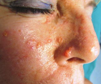

Fig. 14.3
Muir–Torre syndrome. Sebaceous carcinoma on the nose with multiple sebaceous adenomas
Pathology
SC may be classified as well, moderately, or poorly differentiated. The most common presentation is characterized by irregular lobules and sheets with distinctive invasiveness containing cells whose cytoplasm is pale, foamy, and multivacuolated (Figs. 14.4 and 14.5). Some atypical eosinophilic-keratinizing cells with keratin pearls, as seen in squamous cell carcinoma, are found in larger lobules (Fig. 14.6). SC commonly shows considerable nuclear and nucleolar pleomorphism, abnormal mitoses, and zones of central necrosis. In the poorly differentiate variant, basaloid cells are predominant with only a small minority of recognizable sebocytes (Fig. 14.7). Regardless of grade, intraepithelial pagetoid spread and/or multicentric pattern are often seen.
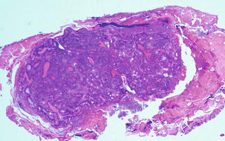
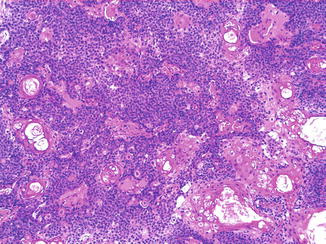
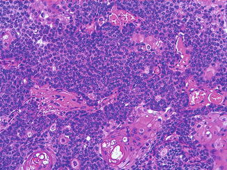

Fig. 14.4
A basaloid dermal-based nodular proliferation of irregular lobules and sheets

Fig. 14.5
Neoplastic cells with basaloid appearance and eosinophilic cells with lipid globules. Some atypical eosinophilic-keratinizing cells with keratin pearls are visible

Fig. 14.6


A basaloid area with scattered vacuolated clear cells showing sebaceous differentiation and keratinizing cells
Stay updated, free articles. Join our Telegram channel

Full access? Get Clinical Tree


