Fig. 14.1
Refractive index. Since the human body contains over 60 % water, this eliminates the need for contrast agent. Therefore, water serves as the standard. The refractive index of water is 1.31. The closer the materials refractive index ratio is to 1.31, the darker the material looks. The further the deviation from this value (+or −), the brighter that material will appear (Reproduced and modified with permission of Caliber I.D. Inc.)
With the use of a near-infrared diode laser, light projected from the confocal microscope focuses on a specific point in the skin. Reflected light from this point is captured through a pinhole-sized opening by a detector as shown in Fig. 14.2. The use of such a small aperture segregates reflected light from the focal section of interest from other areas of the tissue. The depth of light penetration is dependent on wavelength, laser power, reflectivity of the superficial layers of skin, and scattering properties of the dermis. Of all these factors, only laser power can be changed. At longer wavelengths, deeper penetration is gained at the expense of resolution; therefore, present single-laser confocal microscopes are fixed at 830 nm [2].
Fig. 14.2
The basis of the technology: refraction. Confocal scanning laser microscopy technology: after emission, light passes through a beam splitter, a scanning and focusing optical lens, and a skin contact device. Cells and their morphology and cytology are clearly resolved. Diagnostic images are captured at relevant depths in the skin (Reproduced and modified with permission of Caliber I.D. Inc.)
Each section projects as a two-dimensional image (Fig. 14.2) with dimensions of 0.5 mm laterally and 4 mm axially [3] that are oriented parallel (en face) to the skin surface. The resolution of each image is comparable to those of histological sections (Fig. 14.3).

Fig. 14.3
Representative diagram of the skin layers and corresponding RCM images. SC Stratum corneum, SG Stratum granulosum, SS Stratum spinosum, SB Stratum basale, PD Papillary dermis, RD reticular dermis (Reproduced and modified with permission of Caliber I.D. Inc.)
The commercially available near-infrared reflectance microscope (VivaScope 1500, Caliber I.D. Henrietta, NY) is equipped with a diode laser with peak emission at 830 nm and has a maximum power of 35 mW. With this system, each image has an effective 500 × 500 μm field of view and the imaging depth in normal skin is 200–300 μm, i.e., the level of papillary dermis and upper reticular dermis and the spatial resolution in the lateral dimension are 0.5–1.0 μm [4–6]. The new generation of this device (VivaScope 1500, Multilaser) combines reflectance and fluorescence imaging, with available wavelengths of 785 nm (near infrared), 658 nm (red), and 488 nm (blue), which are integrated into a single device amplifying the imaging potentials. These laser wavelengths are applied to the skin, one wavelength at a time, to eliminate the possibility of bleed-through from other fluorophores. The use of several fluorescent dyes has been described including the administration of Indocyanine green (ICG) and fluorescein human skin in vivo [7]. The well-defined intercellular staining pattern of ICG in the skin, with a stain duration of almost 48 hours, has allowed the development of automated cell-recognition algorithms currently under investigation. The expanded capabilities of the multilaser microscope allow the implementation of the device not only in studies seeking the assessment of morphological features in the skin (e.g., atypical honeycomb pattern, atypical keratinocytic and melanocytic nest formation, cell pleomorphism, etc.) but also in research studies aiming to develop targeted fluorescence markers in the field of diagnostic and therapeutic biomarkers, as well as drug delivery studies.
14.2 Clinical Application of RCM as a Diagnostic Tool
A clinical breakthrough in RCM has been the capability to bring to the bedside an almost equivalent tool to histological evaluation without the need for invasive diagnostic procedures. In the setting of cryosurgery, RCM has the capability to aid in the diagnostic decision of a lesion prior to undergoing therapy, which has the advantage of avoiding postponing the treatment until the diagnosis is histologically confirmed. The sensitivity and specificity of RCM varies significantly across tumor types, level of training of the operator, and expertise of the individual interpreting the confocal images [8]. The following skin lesions represent clinical examples in which RCM has been evaluated to date with the potential to offer a diagnostic aid in cases considered for cryosurgery intervention.
14.2.1 Pigmented Lesions
Because melanin and melanocytes are the easiest structures to recognize in RCM, much of the research in the field has been focused on the evaluation of pigmented lesions. RCM has been identified as a useful tool in distinguishing melanomas from benign pigmented lesions (Fig. 14.4a–d). Malignant melanoma in situ (MMIS), lentigo maligna (LM) type, shares similar RCM features such as non-edged papillae, round and large pagetoid cells, and nucleated cells in the dermal papillae with other subtypes of malignant melanoma; however, follicular localization of atypical cells has been proposed as a unique finding in LM. A 2013 systematic review examined five cohort studies on RCM diagnostic accuracy for melanoma, LM, and MMIS in clinically equivocal pigmented lesions using various RCM scoring systems and algorithms [9]. The meta-analysis for these RCM studies demonstrated a per-lesion sensitivity of 93 % and specificity of 76 % for melanoma diagnosis. Thus, RCM features of melanoma may be helpful in noninvasively differentiating equivocal pigmented lesions and delineating margins within lesions such as LM.
Fig. 14.4
(a–d) RCM features in benign and atypical melanocytic lesions. (a) RCM mosaic (4 × 4 mm) acquired at the suprabasal/epidermal-junctional level, showing a ringed and meshwork pattern in a slightly asymmetrical overall architecture. Also milia-like cysts and comedo-like openings are visible throughout the lesion, whereas atypical cells are lacking. The ringed and meshwork pattern fade off at the border of the lesion. (b) Basic image (0.5 × 0.5 mm) showing mostly edged papillae . Arrow is showing the milia-like cyst aspect (→). (c) RCM mosaic (4 × 4 mm) of a very asymmetrical lesion, with a mixed pattern (meshwork, ringed, and clod). In the center of the lesion, a nonhomogeneous meshwork pattern is present, with junctional nests, irregular in size and shape. At the periphery, there are junctional and dermal nests (clods). Dense and sparse and a ringed structure shading off at the periphery. (d) Basic image (0.5 × 0.5 mm) of a central area of the atypical nevus showing dishomogeneous and irregular junctional thickenings and nests surrounding non-edged papillae (red → and *). Within dermal papillae dense and dense and sparse nests are seen (red *), mixed with inflammatory infiltrate, including plump bright cells (yellow *) and small bright spots. Numerous atypical cells are detectable at the DEJ, in single cells or aggregated in nests
Overall, the aid of RCM in the setting of pigmented lesions will be in guiding the clinicians to the areas of greater atypia to perform skin biopsies for histological confirmation and exclusion of invasion [10, 11]. In some cases, invasiveness of melanoma can be readily visualized by RCM, which will aid the clinician when making a decision on whether to perform a cryosurgery intervention or not in an atypical pigmented skin lesion. On the other hand, using RCM as a screening modality for borderline pigmented skin lesions of the head and neck can be of value prior to cryosurgery since, not infrequently, lesions with a lower degree of clinical suspicion can demonstrate increasingly concerning features by dermoscopy and RCM evaluation [12]. This scenario is particularly relevant when evaluating pigmented lesions in chronically photoexposed areas such as the head and neck, as well as distal upper extremity areas [13].
14.2.2 Basal Cell Carcinoma
RCM features of basal cell carcinoma (BCC) have been described at two powers of microscopy: mosaic and individual image levels [14, 15]. At the lower magnification of the mosaic grid, tightly packed tumor cells with high refractility (brightness) can been seen oriented parallel to each other (palisading) at the papillary dermis, forming cord-like structures surrounded by hyporefractile darkness (clefting) that corresponds histologically to the mucinous surrounding stroma. At higher magnification of individual images [14], within the individual tumor cells, elongated hyporefractile nuclei are present, producing a polarizing nuclear appearance [14]. Adjacent to tumor cells, dilated and increased vasculature is often seen with leukocytes on the endothelial lining (Fig. 14.5a–h). Small hyperrefractile dendritic cells within and surrounding the islands of tumor cells have also been identified in pigmented BCCs [15, 16]. A 2004 retrospective study showed that the presence of elongated nuclei and polarized nuclei on RCM was 91.6 % sensitive and 97 % specific for BCC [17].
Fig. 14.5
RCM features in benign and atypical keratinocytic neoplasms. (a) Cobblestone pattern (red arrow) and keratin-filled invaginations . (b) Bright interpapillary spaces (red arrow), melanophages within dermal papillae . (c) Round to triangular, nonnucleated bright cells within dermal papillae (red arrow). Atypical (red arrow) and typical (yellow arrow) honeycomb pattern (HCP). (d) Bright reflectance of the papillary dermis (red arrow). (e) Irregular size of nuclei and thickness of cellular outlines (red arrow); regular HCP (yellow arrow). (f) “Streaming”of spinous keratinocytes (red arrow), regular HCP (yellow arrow) pattern. (g) Tumor islands at the DEJ (red arrow), fibrotic tumor stroma (yellow arrow). (h) Round, oval and trabecular, reflective tumor islands (red arrow); aggregates of melanophages (yellow arrow). All skin lesions shown as macroscopic and corresponding dermoscopy images (10×). RCM mosaic (4 × 4 mm) was obtained for all lesions (Images provided by Curiel and Hofman)
In differentiating between BCC and malignant melanoma (MM), the basaloid cords seen in the papillary dermis are highly specific for BCC [18]. The absence of epidermal honeycomb disarray, papillae (papillae disruption), and cerebriform nests is also useful for diagnosing BCC. Yet, to date, there have been no studies directly comparing pigmented BCCs and MM, and further investigations would be helpful in delineating the RCM features that are unique to each entity. Overall, diagnosis of BCC is one area where RCM holds a high sensitivity and specificity facilitating an RCM diagnostic tool with the potential to spare the need for skin biopsies when interpreted by trained readers.
14.2.3 Actinic Keratosis and Squamous Cell Carcinoma
Actinic keratoses (AK) are one of the most common forms of cutaneous malignancies. While there is debate over whether AK represents a precancerous lesion, studies have shown that without treatment, 0.2–20 % of AKs [19–21] progress to squamous cell carcinomas (SCC). Currently, the diagnostic gold standard for AKs is histopathological examination. While this method represents the most accurate form of identification, it is often time consuming and impractical in daily clinical practice. Over the past six years, a few studies have examined RCM characteristics of clinically diagnosed AK and SCC and their diagnostic value.
Related posts:
 Preoperative Care for Cryosurgery
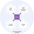 Theoretical Principles of Immunocryosurgery
 Cryobiopsy, Cryoanesthesia, and Cryoanalgesia
 In Vivo Reflectance Confocal Microscopy Assessment of Wound Induction and Repair of a Skin Injury Produced by Liquid Nitrogen: An Atlas
 Cryosurgery for Warts
 Cryosurgery for Vascular Lesions
Stay updated, free articles. Join our Telegram channel

Full access? Get Clinical Tree


