Type of tumor
Preferred type
Benign neoplasia
Infantile hemangioma
Superficial or mixed hemangiomas
Recent appearance, rapid growing, with no signs of involution
Cherry or senile angiomas
Any
Malformations
Hypertrophic capillary malformation
Nodular lesions
For symptomatic and/or cosmetic purpose
Venous malformations
In conjunction with other treatment modalities (surgery, ligation, embolization)
Lymphatic or hemolymphatic malformations
In conjunction with other treatment modalities (surgery, ligation, embolization)
In superficial lesions
For symptomatic and/or cosmetic purpose
Combined vascular malformations
In conjunction with other treatment modalities (surgery, ligation, embolization)
Hyperplasia
Pyogenic granulomas
Any location
Dilation of preexisting vessels
Venous lakes
Depending on vessel size and local blood flow
Spider angioma
Any
Angiokeratomas
Any
In general, cryosurgery is best suited for patients with light skin and for treatment of lesions in non-hair-bearing areas of the body. Among the advantages, one can identify the following: (a) no need of injecting local anesthesia, (b) fast and safe, (c) low cost, and (d) no incision/no suture.
It is a treatment that can be repeated when necessary, and the postoperative care is relatively simple.
12.2.3 When
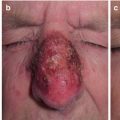
When dealing with vascular tumors, there are multiple noninvasive techniques that will help diagnose and decide which one is the most suitable technique [5]. The first important consideration is to have a diagnostic certainty. For instance, a nasal glioma mimicking a hemangioma [6] or a malignant melanoma can be mistaken for a pyogenic granuloma (Fig. 12.1a, b).
Fig. 12.1
(a) A pyogenic granuloma. (b) A malignant melanoma
12.2.3.1 Dermoscopy
Dermoscopy can be used for diagnostic purposes [7] as well as a follow-up to cryosurgical treatment. A dermoscopic picture of the lesion can be compared to the postsurgical image in order to determine the improvement by verifying the reduction in lesion or in vessels (Fig. 12.2).
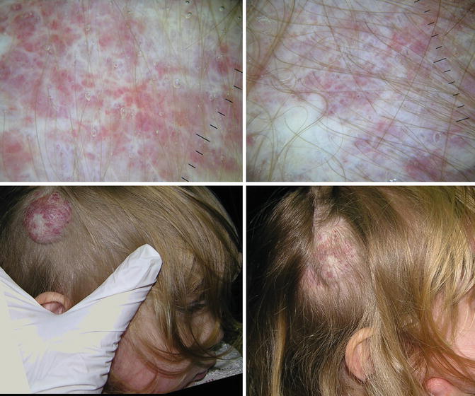
Fig. 12.2
A dermoscopic image of a capillary hemangioma taken before treatment (left column) and at control evaluation 1 month later. It puts into evidence the areas with fibrous tissue, the diminished number of capillaries, and the color difference
12.2.3.2 Temperature Control
The surface temperature of a vascular lesion is usually higher by several degrees than body temperature. By measuring the tumor’s surface temperature before and after treatment, one can register improvement. After treatment, there should be fewer vessels present and therefore a lower temperature. An infrared technology or a temple touch thermometer will be sufficient to monitor the tissue temperature (Fig. 12.3).
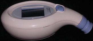
Fig. 12.3
A temple touch thermometer used to measure superficial temperature in capillary hemangiomas (Courtesy of and Copyright © 2014 Medline Industries, Inc.)
12.2.3.3 High-Frequency Ultrasound/Doppler
Ultrasound (US), especially with a high-resolution transducer, has been advocated as useful in examining small and superficial soft tissue masses that are suggestive of hemangiomas or vascular malformations. Vascular tumor ultrasound stands as one of the easier and more universally practiced noninvasive techniques [8] that give information about size, shape, depth, and, more importantly, vascularity [9]. In addition, US will help differentiate between a cystic and a solid lesion. Doppler US will give information about the presence or absence of flow and the degree of vascularization (Fig. 12.4). The Doppler characteristics of vascular malformations are helpful in differentiating low- from high-flow vascular malformations [10]. The sonographic depiction of abundant low-flow vascular channels can be a predictor of the potential success of percutaneous sclerosis. In addition, US can be used to guide needle placement during percutaneous sclerosis in a combination treatment approach (cryosurgery plus infiltration).
Fig. 12.4
Color Doppler analysis of the vascular mapping of a vascular malformation in the abdomen of a child
12.2.3.4 CT Scan Angiography and Nuclear Magnetic Resonance Imaging
CT scan angiography is an imaging technique that combines a CT scan with an injection of contrast material to produce a picture of the vasculature of a tumor. It can help differentiate between a hemangioma and a vascular malformation [11]. A 3D reconstruction displays the total volume of the lesion as well as the thickness and number of vessels (Fig. 12.5). MR imaging with gadolinium contrast is the diagnostic modality of choice for large and deep lesions or for those where US has not been diagnostic [8]. Intravenous gadolinium helps differentiate between cystic or solid tumors and between venous and lymphatic malformations [12], allowing the determining of lesion size and number and size of vessels involved [13]. This information is necessary to determine the feasibility of cryosurgical treatment or the need to combine treatment modalities.
Fig. 12.5
3D reconstruction of a CT scan done on a mixed capillary hemangioma in the frontal area of the same patient in Fig. 12.6
12.2.3.5 Other Noninvasive Imaging Techniques: Reflectance Confocal Microscopy (RCM)
Confocal microscopy might be helpful in characterizing vascular lesions in vivo before cryosurgical treatment. There is good correlation between RCM morphology and established histopathology criteria for common vascular lesions such as pyogenic granuloma, capillary vascular malformations, and venous lakes [14].
12.2.4 How
12.2.4.1 The Rule of the 4 Ps
As a general rule for most vascular lesions, cryosurgical treatment is performed with probe or close technique, although there are conditions where spraying or tweezers can work as well. When using probes, there is a simple rule to follow called the 4P rule (probe, pressure, previously frozen probe, patience):
Probe: Blood is a warm liquid (around 38 ºC) that has a slightly higher temperature than the body. If a superficial freezing is performed, there will only be destruction of the most superficial skin structures, leaving the rest of the vessels untreated. Blood present in deeply seated vessels will keep the area warm. Therefore, the ideal treatment of vascular lesions is with probes (contact or close cryosurgery). Probes will freeze deeper at a constant low temperature. Metal probes limit the “heat sink” effect.
Pressure: Vascular tumors tend to be non-painful renitent tumors. They can be pressed, squeezing out some of the blood inside them. By doing so, the lesion’s temperature goes down (less blood, less temperature). Pressure minimizes blood flow. As the blood flow is reduced, local temperature will be lower and cryosurgical treatment more effective.
Previously frozen probe: A previously frozen probe sticks less to the surface. When treating vascular tumors, it is best to have the possibility of removing it fast from the skin surface. The reasons are (a) a sudden movement while doing the procedure from the patient or surgeon’s part could cause an accidental breaking of the vascular tissue and a profuse undesired bleeding (this is especially true when dealing with small children). (b) Once freezing has been completed, the probe has to be removed as soon as possible to avoid further freezing that could leave an undesired scar. In vascular tumors, there is no need to freeze margins above 1 mm.
Patience: Despite all precautions such as drying the surface and previously freezing the probe, probes can occasionally get stuck to the surface. Be patient: the need to remove the probe from the site should not tempt the surgeon to force it out because this will simply break the tissue. In the case of a vascular lesion, bleeding can become a real issue. If, in spite of these precautions, the probe cannot be removed, allowing some warm water to run over the probe will rapidly release it from the surface. Keep a cup with warm water at hand as a precaution at all times.
12.3 Capillary Hemangiomas
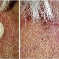
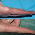
Capillary hemangiomas, also called strawberry or infantile hemangiomas, are sometimes classified as benign neoplasms or vascular hyperplasias. They are the most frequent benign tumors of infancy and are composed of proliferating endothelial-like cells. Clinically, they can present themselves as superficial, deep, or mixed hemangioma (Fig. 12.6a, b), with regard to the depth of the vascular nest within the skin. Superficial hemangioma are bright red and sometimes called strawberry tumors. Deep lesions are violaceous tumors covered with normal skin, and mixed lesions have the bright and granulous surface but tend to be elevated. Histopathologically, hemangiomas can present as well-circumscribed, densely packed endothelial cells and small vascular spaces. These tumors usually appear after 1–2 weeks of birth and begin a rapid process of growth that can last up to 6 months. They sometimes ulcerate in the process of growing. After the growth period, they begin a very slow involution period that can last up to 6–7 years. During this period, certain hemangiomas ulcerate. Therefore, ulceration can be a sign of both rapid growth or involution [15].
Fig. 12.6
(a) A large frontal mixed capillary hemangioma ulcerated in the center. A combined treatment with steroid infiltration for the deep part of the lesion and close (probe) technique on the ulcerated superficial and medial part of the tumor. (b) A control image 3 years after treatment
According to the American Academy of Dermatology, the goals in the management of hemangiomas are to prevent or reverse the complication of alarming hemangiomas, prevent permanent disfigurement, minimize psychosocial stress for patients and relatives, avoid aggressive and potentially unsightly procedures, and prevent or adequately treat ulceration, thereby minimizing scarring, infections, and pain [16].
Possible interventions are left for those rapidly growing hemangiomas, especially those in areas that put the child in danger, such as:
Tumors close to airways that can limit breathing capacity.
Bleeding tumors.Related posts:

Stay updated, free articles. Join our Telegram channel

Full access? Get Clinical Tree