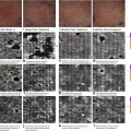
Fig. 26.1
Two confocal images obtained from the same skin site and from the same depth (approximately 80 μm) on Day 1 (a) and on Day 3 (b). Papillae and some keratinocytes loaded with melanin granules can be seen
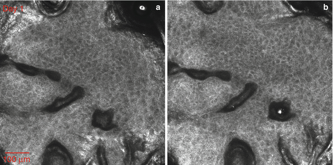
Fig. 26.2
Two confocal images obtained at the same depth of the stratum granulosum on Day 1 (a) and Day 5 (b). The scale is the same as in Fig. 26.1a, b. We show the alignment of skin sites at the level of the granulocytes. In this case the undulations of the surface of the stratum corneum and sweat glands as well as hair follicles play a role in the alignment of images. It is difficult to follow individual cells in these images or their migration from day to day although the alignment would cause us to assume that it might be possible
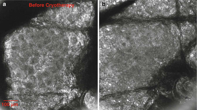
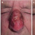
Fig. 26.3
Imaging the same location on the skin allows us to study morphological changes after cryotherapy. The two images were captured just below the stratum corneum on Day 1 (a) and Day 3 (b). The cell outlines in the image on the left correspond to keratinocytes within the granular layer. Post-injury, cellular membranes lost definition and glyphic islands flattened. What we found to be surprising in the confocal images immediately after freezing was that the cell membranes in the stratum granulosum and stratum spinosum remained intact contrary to the assumption that the cells rupture upon freezing because of the expansion of ice crystals as the temperature is lowered. The cell outlines became imperceptible at Day1 after freezing
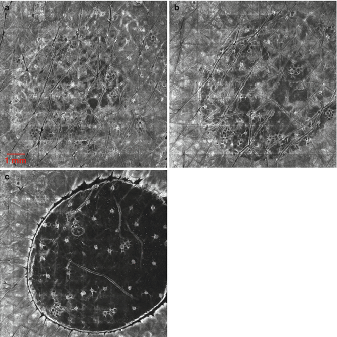
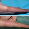
Fig. 26.4
Each of the microimages is a composite of 10 × 10 individual microscopic fields (0.5 × 0.5 mm). The area that was frozen is distinguishable in each image. The three images were obtained from the same site on Days 1 (a), 3 (b), and 7 (c) after freezing. Depth of the image mosaics is 70 μm below the surface of the stratum corneum. Soon after freezing a blister forms due to the separation of the epidermis from the dermis. Evidence of the blister may be documented from Day 1 after freezing and becomes fully separated by Day 2 to Day 3. The papillae may be seen in the image from Day 1, and they become imperceptible as full separation is achieved
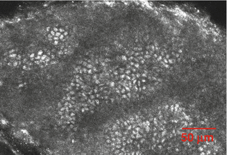
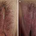
Fig. 26.5


A confocal image obtained from within the blister roof – the damaged epidermis – on Day 7 after freezing with liquid nitrogen. The melanin granules at or close to where the basal layer was remain intact and have not diffused beyond the original cellular boundaries. There is no longer evidence of cell boundaries. As full epidermal separation occurred, the sweat glands remain intact, connecting the dead epidermis to the underlying dermis with blister fluid in between the two which serves as a good optical coupling medium. The epidermis of lentigines is found to contain a large number of refractive cells believed to be bright because of scattering by the melanin granules in the melanin-bearing keratinocytes. We were interested to find out the extent to which the melanin granules would migrate to the intercellular space once the keratinocyte membranes were ruptured. There was no redistribution of melanin granules following the disappearance of the cell membranes. This finding was consistent for all volunteers with lentigines
Related posts:


Stay updated, free articles. Join our Telegram channel
Full access? Get Clinical Tree
