19 Rheumatologic conditions of the hand and wrist
Synopsis


Introduction
Rheumatoid arthritis (RA) is a systemic autoimmune disease that affects 1% of adults (ranging from 0.33% to 6.8%, depending upon the population).1 Although RA has many diverse clinical manifestations, the single underlying pathologic process is synovial inflammation, which progressively destroys joints and soft tissue leading to deformity and disability. Over the last 20 years, many advances in the medical management of RA have been made. New medications have improved the ability to control rheumatoid inflammation, and can dramatically slow or prevent disease progression.2 In spite of these tremendous strides, the hand surgeon continues to play a crucial role in treating RA. Many patients continue to suffer from recalcitrant pain or disability despite maximal medical management, and may benefit from surgical intervention.
Basic science/disease process
Etiology
Though much is understood about the pathogenesis of RA, the cause remains idiopathic. That being said, RA is known to have a genetic component. This is demonstrated by the fact that RA has a much higher concurrence rate in monozygotic twins (15–20%) than in dizygotic twins (5%), with susceptibility transferred in an autosomal recessive mode.3–5 In addition, there are certain ethnic groups such as the Pima Indians that exhibit a greater incidence of RA than the rest of the population, providing further evidence of a genetic component to RA.6
Many studies suggest that a multitude of genes play a role in contributing to susceptibility to RA. These include the class II major histocompatibility complex (MHC) genes and many others. The most well-understood genetic association is with the class II MHC genes, which may contribute up to 40% of the genetic component of RA.7 One class II MHC gene in particular, human leukocyte antigen (HLA) DR4 is associated not only with increased risk of disease, but also with increased disease severity.4
In addition to genetics, gender influences the development of RA. RA is more common in women than in men, with a ratio of 2 : 1 to 3 : 1. Multiple laboratory and clinical studies suggest that hormones, and estrogens in particular, affect the development of RA. However, the exact role of estrogen in the development of RA is not known.8
Although the mechanisms are not well understood, the environment plays a role in the etiology of RA as well. Smoking is a risk factor for the development of RA, particularly in susceptible men.9 Coffee intake and exposure to silica have also been associated with RA.4 In addition, infectious diseases likely act as a trigger in genetically susceptible individuals. Possible triggering agents include mycoplasma, enteric bacteria, Epstein–Barr virus, and others viral and bacterial triggers.10–13 In summary, RA is an idiopathic disease, but which is known to have both genetic and environmental components. The genetic susceptibility and environmental triggers are complex and varied, and are not fully understood.
Pathogenesis
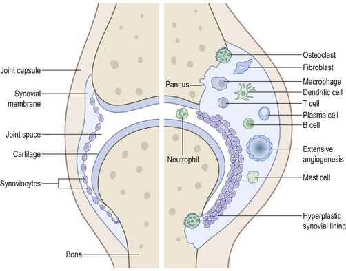
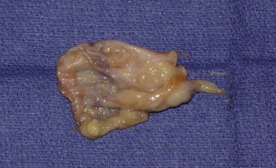
The target tissue of RA is the synovium. The disease process begins when an antigen, a trigger for RA, is presented to T-cells within the synovium. A complex interaction between macrophages, B-cells, T-cells, and synoviocytes ensues, orchestrated by systemic inflammatory modulators and local cytokines. The end result is the formation of proliferating inflamed synovium, or pannus (Figs 19.1, 19.2). Once initiated, this process becomes systemic and self-sustaining, and does not require the prolonged presence of the trigger antigen. The synovial pannus produces proteolytic enzymes such as metalloproteinases, serine proteases, cathepsins, and aggrecanases that erode cartilage, bone, and supporting soft tissue structures. Cytokines secreted by the pannus activate osteoclasts in nearby bone, further contributing to bony erosions and joint destruction.14–16
Medical management
The conventional DMARDs include methotrexate, leflunomide (Arava), azathioprine (Imuran), Plaquenil, and gold.17 Methotrexate is the most commonly administered DMARD because of its effectiveness and favorable side-effect profile. It is generally taken orally once a week, in combination with folic acid in order to minimize side effects such as hepatotoxicity and bone-marrow suppression. Leflunomide (Arava) is a daily oral pyrimidine antagonist, with side-effects that include hepatotoxicity and marrow suppression. Plaquenil, an antimalarial drug that is effective in the treatment of RA, is taken daily and has few serious side-effects. Sulfasalazine is another common daily DMARD, and like many of the other conventional DMARDs it can cause leukopenia. Gold is rarely included in the antirheumatic regimen today due to significant side-effects and the need for intramuscular injections.
Biologic DMARDs are those that target tumor necrosis factor alpha (TNF-α) and interleukin 1 (IL-1), or the cellular components of the immune system. Etanercept (Enbrel) is an anti-TNF-α that requires once-weekly subcutaneous injections. Infliximab (Remicade) is a TNF-α blocker that is administered every 1–2 months, intravenously. Adalimumab (Humira) is a TNF-α blocker that is injected subcutaneously every two weeks. All three are very effective in treating RA, especially when combined with methotrexate.18,19 However, the long-term toxicity of these new medications is unknown, and all three result in an increased susceptibility to infections.20 Anakinra (Kineret) is a biologic DMARD that blocks IL-1 receptors, and requires daily subcutaneous injections. Like the anti-TNF-α biologics, it is associated with an increased susceptibility to infections. Rituximab (Rituxan) is an intravenously administered monoclonal antibody that targets certain B-cells, and lasts from many months up to a year. Abatacept (Orencia) is an antibody that targets T-cells, and requires a monthly intravenous injection.
The perioperative management of DMARDs and other rheumatoid medications is somewhat controversial and should be coordinated with the patient’s rheumatologist. The perioperative cessation of DMARDs or corticosteroids can result in an acute deterioration (rheumatoid flare-up) that is poorly tolerated by the patient. In fact, the flare-up may be so severe that it affects the patient’s ability to participate in postoperative rehabilitation.21–23 In general, methotrexate should be continued throughout the perioperative period at its normal dose. Methotrexate does not appear to increase infection rates in patients with RA undergoing elective surgery.21,22 Furthermore, corticosteroids, when used alone or in combination with methotrexate, do not affect wound infection rates and should not be stopped in the perioperative period.23 Data on the other conventional DMARDs are more limited, and their perioperative dosing should be discussed with the patient’s rheumatologist.
The appropriate perioperative management of biologic DMARDs, particularly the TNF-α inhibitors, is less clear. There are few data and no large studies to guide perioperative dosing of the biologic DMARDs.22 Therefore, a conservative approach is recommended at this time. In general, the TNF-α inhibitors should be held 2–4 weeks before and after surgery.22 Similarly, few data exist regarding the other biologics, and a similar approach is recommended.24
Hints and tips
Perioperative management of anti-rheumatic medications
| Medication | Recommendation |
|---|---|
| Corticosteroids | Continue at normal preoperative dosage Provide stress-dose at time of surgery if taking >5–10 mg/day, or if recently stopped long-term corticosteroid use |
| Methotrexate | Continue at normal preoperative dosage |
| Other conventional DMARDs | Discuss with rheumatologist, but in general continue |
| Biologic DMARDs | Discuss with rheumatologist, but in general hold for 2–4 weeks before and after surgery |
Diagnosis/presentation
The onset of RA is usually in the 3rd to 6th decade of life. The incidence of RA in young men is one-third that of women, but increases to be equal to that of women by the 6th or 7th decades of life. From the onset, RA tends to affect the hands and wrists. In fact, the metacarpophalangeal (MCP) joints, proximal interphalangeal (PIP) joints, and the wrist are often affected earlier and more frequently than other joints in the body.2
Diagnostic criteria have been described, and are useful in making the diagnosis of RA. They include: (1) morning stiffness = 1 h; (2) soft tissue swelling of three or more joints; (3) soft tissue swelling of hand joints (PIP, MCP, wrist); (4) symmetrical soft tissue swelling; (5) subcutaneous nodules (rheumatoid nodules); (6) seropositivity for rheumatoid factor (RF), and (7) erosions or periarticular osteopenia in hand or wrist joints. Four of the seven criteria should be present for diagnosis, and criteria (1) through (4) must be present for 6 weeks or longer. Morning stiffness, or stiffness after rest is common, and may be present for a number of hours, improving with hand use. The most sensitive findings are symmetric arthritis and arthritis of the hand joints. Classic radiographic changes, and the presence of rheumatoid nodules are less sensitive but very specific findings. RF is not only is a marker for RA, but also correlates with disease severity.4 Although not one of the classification criteria, anticitrullinated protein antibodies (ACPAs) are very specific for RA, and are useful in confirming the diagnosis.4,25 Like RF, ACPA seropositivity also correlates with disease severity.26 In many cases, seropositivity to RF and ACPA develops prior to the onset of clinical disease. It should be noted however, that some patients with RA will convert to seropositivity after the onset of disease, and some patients with RA will never become seropositive.
RA usually develops in multiple joints slowly over a period of months, although atypical presentations are not unusual.27 In some cases, symptoms can occur rapidly over a few days. When symptoms develop rapidly, the diagnosis of RA is often not considered, and septic arthritis or some other cause of acute-onset joint inflammation is initially suspected. In other cases, symptoms may involve one or two joints for some time before becoming polyarticular, confounding the diagnosis. Less commonly, some patients develop extra-articular disease (rheumatoid nodules, vasculitis, pericarditis, pleural effusion or interstitial disease, peripheral neuropathy, keratoconjunctivitis sicca, and many others), before synovitis and arthritis are evident.4 Palindromic rheumatism is a rare variant of RA, in which symptoms begin in a single joint. Symptoms worsen and spread to other joints for a few days, then disappear in reverse order. Although the disease resolves in some patients, about half eventually develop classic RA.28 Finally, it has been noted that some patients tolerate RA quite well; a presentation termed arthritis robustus. This tends to occur in athletes or heavy laborers who have little pain or disability in spite of severe radiographic changes.
Any joint in the body can be affected by RA, including the articulations of the ossicles of the middle ear.29,4 However, joint involvement tends to follow a predictable pattern. Symptoms often begin in the MCP, PIP and wrist joints first, in part due to their high synovium to joint surface area ratio.30 Larger joints like the knees, hips, shoulders and elbows are likely to be affected later in the disease process due to their lower synovium to joint surface area ratio.31 Within the hand, the DIP joints are generally spared, possibly because of their relatively small absolute amount of synovium. The synovial lining of tendons is also affected by RA, and can result in a multitude of symptoms, including carpal tunnel syndrome, tendon rupture, tendonitis, and trigger digits.
It should be remembered that RA is a systemic disease with multiple extra-articular manifestations. Rheumatoid nodules, vasculitis, pericarditis, pleural effusion, interstitial pulmonary disease, peripheral neuropathy, and keratoconjunctivitis sicca are some of the more common extra-articular effects of RA. Although rheumatoid nodules are present in only a minority of patients with RA, they are a common reason for presentation to the hand surgeon. Rheumatoid nodules develop secondary to the same autoimmune process that occurs in the joints. They occur most often on extensor surfaces or areas of contact pressure such as the olecranon process. However, they can appear anywhere, including within internal organs such as the lungs, heart, or central nervous system.32
Hints and tips
Diagnostic criteria for rheumatoid arthritis
| Criterion | Details |
|---|---|
| 1. Morning stiffness | 1 h |
| 2. Soft tissue swelling | 3 or more joints |
| 3. Soft tissue swelling | Symmetric |
| 4. Soft tissue swelling of hand | MCP, PIP, wrist joints |
| 5. Subcutaneous nodules | Rheumatoid nodules |
| 6. Seropositivity | Rheumatoid factor (RF) |
| 7. Typical radiographic findings | Periarticular erosions/osteopenia, hand or wrist |
Four of the seven criteria must be present, and criteria 1–4 must be present for at least 6 weeks.
Wrist involvement
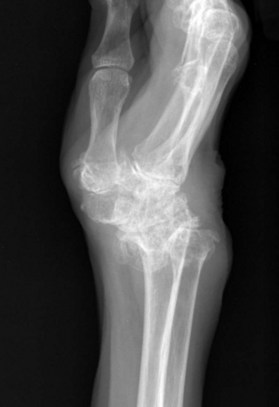
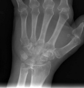
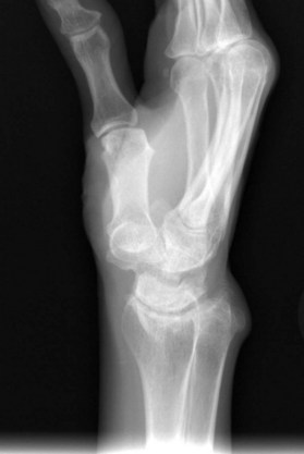
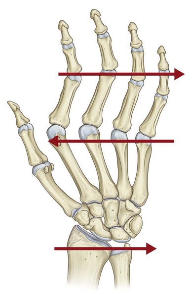
Within the wrist and distal radioulnar joint (DRUJ), cartilage is degraded and bony erosions develop. Bony erosions occur first at sites of nutrient vessels and at the joint margins because there is no protective cartilage at these locations, thereby giving the invading pannus direct access to the bone.33 Radiographic changes are first noted at the scaphoid waist, the ulnar styloid and the distal radioulnar joint (DRUJ) (Fig. 19.3).34,35 As the arthritis progresses, the radiocarpal joint becomes involved. Bony destruction tends to be more severe at the radiocarpal joint than the mid-carpal joint, although both joints are often involved.36 In late stages, the volar lip of the distal radius becomes severely eroded, allowing proximal migration, volar translation and volar angulation of the lunate, with compensatory mid-carpal extension (Figs 19.4, 19.5).35,37


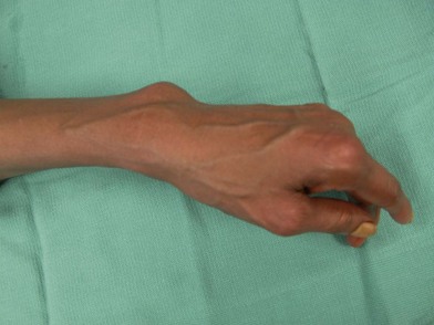
The ligaments of the wrist and DRUJ are affected as well. As the pannus proliferates, it stretches and then directly invades the intrinsic and extrinsic wrist ligaments. Synovitis occurring at the scaphoid waist weakens the radioscaphocapitate ligament, leading to ulnar translocation of the carpus (Fig. 19.6).38 Destruction of the scapholunate ligament leads to carpal instability, and further contributes to ulnar translocation of the carpus. At the DRUJ, pannus invades the triangular fibrocartilage complex (TFCC), including the dorsal and palmar radioulnar ligaments, resulting in DRUJ instability and eventually dorsal dislocation of the ulnar head. Pannus also destroys the extensor carpi ulnaris (ECU) subsheath, resulting in volar subluxation of the ECU.39,40 The ECU’s function as a wrist extensor and ulnar deviator is lost, contributing to supination and radial deviation of the carpus. The combination of dorsal dislocation of the ulnar head, carpal supination and volar subluxation of the ECU is termed the “caput ulnae” (Figs 19.7, 19.8).
End-stage wrist arthritis falls into three different clinical patterns, depending upon the degree of bony destruction and ligamentous instability.41 These patterns are: (1) ankylosis; (2) arthritic stable, and (3) unstable (subdivided into ligamentous unstable and bony unstable). In the ankylosing wrist, the carpus undergoes autofusion. Frequently, ankylosis occurs with the wrist in acceptable alignment, although this is not always the case. In the arthritic stable wrist, the arthritis follows a pattern similar to that of osteoarthritis. Ligamentous destruction is limited, and the wrist remains stable over time. In the unstable wrist, there is progressive malalignment. In the ligamentous unstable wrist, bony destruction is minimal, whereas in the bony unstable wrist, there is severe loss of bone resulting in instability and eventually dislocation.
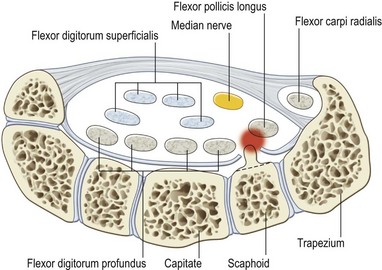
On the dorsal aspect of the wrist, synovitis develops within the extensor tendon compartments (Fig. 19.9). Depending upon the extent of the synovitis, there may be a bulge protruding beyond the distal and proximal margins of the extensor retinaculum, creating an hourglass appearance. Direct invasion of extensor tendons, combined with attritional wear over sharp bony edges at the ulna and DRUJ can lead to tendon ruptures. The extensor tendons to the small finger are usually affected first. Extensor tendon ruptures often progress radially, eventually affecting all digits. This ulnar to radial progression of extensor tendon ruptures is called the Vaughn–Jackson syndrome (Fig. 19.10).42 Tendon ruptures are sudden, and may not be painful. They can be difficult to identify in patients with severe deformity, and must be differentiated from volar subluxation of the MCP joint, radial nerve palsy, and extensor tendon subluxation at the MCP joint. On the volar aspect of the wrist, sharp erosions on the scaphoid can result in attritional rupture of the flexor pollicis longus (FPL) tendon, known as a Mannerfelt lesion (Fig. 19.11).43 Within the carpal tunnel, synovial pannus and tenosynovitis proliferate, creating a space-occupying lesion that results in carpal tunnel syndrome (Fig. 19.12).
Finger and thumb involvement
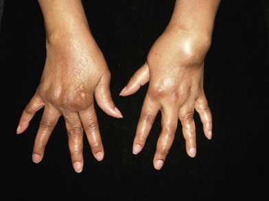
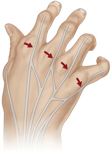
At the MCP joints, the typical pattern of instability is volar subluxation and ulnar deviation, and is the result of multiple forces.44,45 Synovitis at the MCP joint erodes the dorsoradial portion of the joint capsule early in the disease process, creating an early tendency towards ulnar deviation (Fig. 19.13) and volar subluxation (Fig. 19.14).46,47 Radial deviation at the wrist creates an ulnar approach of the extensor tendons to the MCP joints, which further contributes to ulnar deviation (Fig. 19.15).48 In addition, ulnar deviation forces during oppositional and key pinch stretch the dorsoradial portion of the capsule and the radial collateral ligament, particularly in the index and long fingers.49 Finally, continued dorsoradial invasion of the pannus weakens the radial sagittal bands, causing ulnar subluxation of the extensor tendons between the metacarpal heads, which further increases ulnar deviation force on the MCP joints, and weakens extension power at the MCP joints (Fig. 19.16).35,50
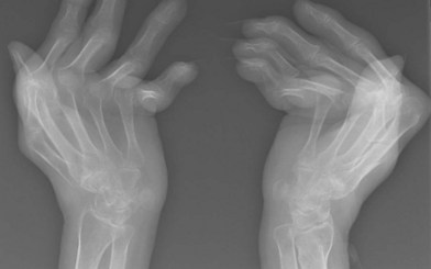
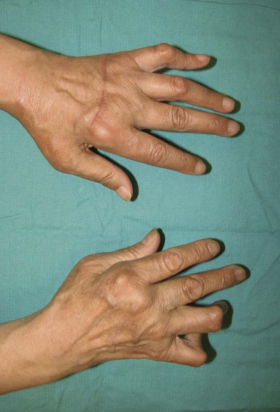
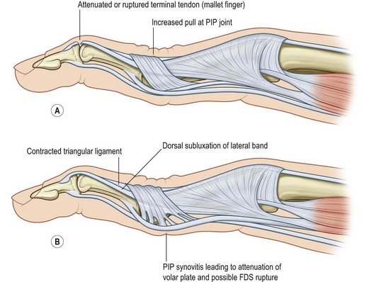
Fig. 19.14 Radiograph of bilateral hands, demonstrating flexion and volar subluxation of the MCP joints.
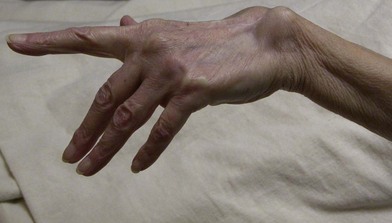
One of two patterns of finger deformity can occur, the swan-neck (Fig. 19.17) or boutonniere deformity (Fig. 19.18). Although swan-neck deformity is more common, either deformity can occur and both can occur in the same hand. Swan-neck deformity can originate from pathology at the MCP joint, the PIP joint, or the DIP joint (Fig. 19.19). At the DIP joint the terminal tendon insertion attenuates or erodes, resulting in a mallet finger. This causes an imbalance in the extensor mechanism, with increased extension force at the PIP joint. MCP flexion also contributes to increased pull of the central slip at the PIP joint. When these increased extension forces are combined with synovial disruption of the PIP volar plate, PIP hyperextension occurs, resulting in swan-neck deformity. Swan-neck deformity can originate at the PIP joint as well. Pannus stretches and erodes the volar plate and capsule, allowing hyperextension. Flexor tendon rupture may also contribute to a loss of flexion force at the PIP joint. The lateral bands slide dorsally, limiting PIP flexion. DIP joint flexion is a secondary phenomenon in this scenario, and is due to slack in the extensor mechanism combined with tightening of the flexor digitorum profundus (FDP) from PIP hyperextension. MCP pathology can initiate the development of swan-neck deformity as well. Flexed MCP position results in excessive pull through the extensor mechanism. The intrinsics also tighten over time, thereby hyperextending the PIP joint when MCP extension is attempted. The lateral bands also migrate dorsally, contributing to the deformity. The most debilitating result of swan-neck deformities is loss of flexion at the PIP joints, resulting in an inability to flex the fingers for pinching and grasping.
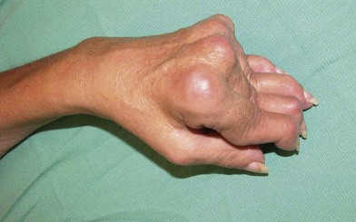
Fig. 19.17 Classic swan-neck deformities of all fingers, with MCP flexion, PIP hyperextension, and DIP flexion.
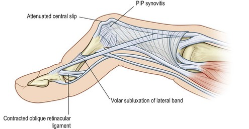
Boutonniere deformities are usually less debilitating than swan-neck deformities, because pinch and grasp are usually preserved. Unlike swan-neck deformities, boutonniere deformities always begin with pathology at the PIP joint (Fig. 19.20). The initial insult is stretching, erosion, and rupture of the central slip insertion on the base of the middle phalanx. Attenuation of the dorsal capsule, transverse retinacular ligament and triangular ligament allows the lateral bands to sublux volar to the joint axis of rotation, becoming PIP flexors. The volar position of the lateral bands makes them even more effective extensors of the DIP joint, resulting in secondary DIP hyperextension.
Synovitis also develops within the flexor tendon sheath, and can lead to triggering or rupture of the flexor tendons. Trigger digits associated with RA are distinct in mechanism and pathology from nonrheumatoid trigger digits, and occur secondary to synovitis or small rheumatoid nodules in the flexor tendon.51 The specific location of the synovitis or rheumatoid nodules determines the presentation of the rheumatoid trigger finger. A nodule located proximal to the A1 pulley may present like a typical nonrheumatoid trigger finger with locking in flexion, or triggering during extension. A nodule just distal to the A2 pulley can result in the finger locking in extension, or triggering during flexion. Diffuse tenosynovitis or multiple nodules can result in swelling and loss of flexion and extension.52,53
Thumb deformity can be organized into five categories. Type I, the most common, is a boutonniere deformity with MCP flexion, IP hyperextension, and radial abduction of the metacarpal (Fig. 19.21). Deformity begins when synovial pannus at the MCP joint erodes dorsally, attenuating and eventually rupturing the extensor pollicis brevis (EPB) tendon insertion, and displacing the extensor pollicis longus (EPL) ulnarly and volarly. Because of the loss of dorsal capsule and the loss of the EPB, the MCP joint flexes and subluxates volarly. Hyperextension at the IP joint occurs secondarily, and can be exacerbated in patients with an FPL rupture. Type III rheumatoid thumb deformity is a swan-neck deformity, and is the second most common deformity, presenting with MCP hyperextension, IP flexion, and metacarpal adduction contracture. This deformity begins at the carpometacarpal (CMC) joint, with attenuation of the volar beak ligament. As the CMC joint subluxates or dislocates, metacarpal adduction occurs in a similar fashion to nonrheumatoid CMC arthritis. MCP joint hyperextension occurs secondarily as the thumb compensates for the adducted metacarpal, and is exacerbated by volar plate attenuation or erosion from pannus invasion.
Patient selection
Perioperative considerations
Care must be taken during the preoperative workup of RA patients, because many unique problems can impact the safety and timing of surgery. Airway management can be quite difficult. Temporomandibular joint arthritis can limit oral opening, making intubation difficult. Glottic narrowing can also occur due to inflammation or arthritis of the cricoarytenoid joints, creating further difficulty with airway management.54 Atlantoaxial instability is another concern, and is quite common in patients with RA. Flexion of the neck during intubation in patients with significant instability can cause spinal cord injury or death. Therefore, preoperative cervical spine flexion-extension radiographs should be obtained in all rheumatoid patients preoperatively. Fiberoptic intubation with the patient in a cervical collar is recommended in patients with atlantoaxial instability.55
Treatment/surgical technique
Operations at the wrist
Wrist synovectomy/dorsal tenosynovectomy
A minority of patients will have persistent wrist synovitis or dorsal tenosynovitis that is painful and unresponsive to at least 6 months of maximal medical treatment. Wrist synovectomy and/or dorsal tenosynovectomy may be effective in improving pain in these patients.56,57 Whether this significantly slows the progression of disease is unknown.
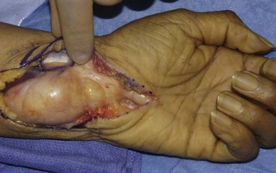
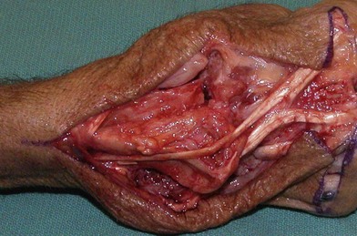
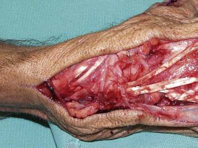
A midline longitudinal incision is made over the dorsal wrist, and skin flaps are elevated at the level of the extensor retinaculum, preserving the radial and ulnar sensory nerves that lie within the subcutaneous tissue. If there is a large amount of synovitis, it can be seen deep to the attenuated extensor retinaculum (Fig. 19.22). The extensor retinaculum is incised along the radial border of the first extensor compartment, leaving enough substance intact for later closure. The retinaculum is elevated in an ulnar direction to, but not into the sixth extensor compartment, exposing the extensor tendons. The extensor retinaculum is often very attenuated, and care should be taken to preserve its integrity during elevation. Extensor tenosynovectomy is then performed. Working systematically, each extensor tendon is retracted individually and synovium is excised with curved tenotomy scissors (Fig. 19.23). In some cases, it becomes clear during tenosynovectomy that an extensor tendon rupture has previously occurred, and that the ruptured extensor tendon or tendons are held together by a mass of adhesions and synovial pannus. This situation should be discussed preoperatively with the patient, as tendon transfers will be required. After completing the extensor tenosynovectomy, a posterior interosseous neurectomy may be performed to improve pain relief. The posterior interosseous nerve is identified on the floor of the fourth extensor compartment and a 2 cm segment is excised, with cauterization of the proximal stump.
Fig. 19.22 Extensor tenosynovitis of the right wrist, with severely attenuated extensor retinaculum.
If any rough bony surfaces remain exposed after closure of the capsule, the retinaculum can be divided transversely, creating two ulnarly based retinacular flaps. One flap is sutured deep to the extensor tendons, covering the exposed bony surfaces. The other retinacular flap is closed dorsal to the extensor tendons, leaving the EPL transposed (Fig. 19.24). Alternatively, if the ECU tendon is subluxated volarly, the retinaculum can be used to stabilize it. The ECU tendon is synovectomized and relocated to its normal position dorsal to the ulna. Half of the retinacular flap is placed deep to the ECU tendon, then wrapped back dorsally and radially and sutured to itself at the border of the fifth extensor compartment, securing the ECU dorsal to the wrist axis of rotation.
Distal ulna resection (Darrach procedure)
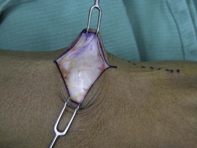
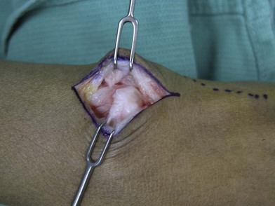
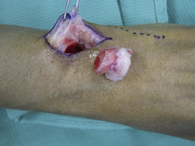
If performed in isolation, a 3 cm longitudinally oriented chevron incision is made over the dorsal aspect of the ulnar head (Fig. 19.25). If combined with another wrist procedure, skin flaps and retinacular flaps are elevated, and extensor tenosynovectomies are performed as described above. A longitudinal incision is made in the floor of the fifth extensor compartment, with a 90° extension ulnarly just distal to the head of the ulna, and just proximal to the TFCC. Although it is often destroyed, when the TFCC is present, it should be preserved. The capsule is elevated off the ulnar head, and the ulnar head and neck are dissected subperiosteally with a 15-blade and Freer elevator (Fig. 19.26). The ECU is usually subluxed volarly, and is elevated off the ulna during subperiosteal dissection. Hohmann retractors are used to provide exposure of the neck and head of the ulna. Once the ulnar head and neck have been exposed circumferentially, a sagittal saw is used to create a transverse osteotomy at the neck of the ulna, at the level of the proximal margin of the sigmoid notch of the radius. Any remaining soft tissue attachments are released, and the ulnar head is removed (Fig. 19.27). The dorsal edge of the ulnar stump is then beveled with the sagittal saw, and rasped smooth to prevent tendon abrasion and rupture. Any remaining synovial pannus can now be easily accessed, and is debrided with a synovial rongeur.
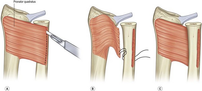
Prior to closure, the ulnar stump should be stabilized and the normal relationship between the ulna and the carpus should be re-established or improved. The pronator quadratus is elevated subperiosteally from the volar ulnar aspect of the ulna, leaving its attachment to the radius intact. It is important to keep the periosteum attached to the pronator quadratus. It is then delivered through the interosseous space and draped dorsally over the ulnar stump. The ulna is reduced volarly. While maintaining the ulnar stump in reduction, the pronator quadratus and its periosteum are sutured securely to the dorsal aspect of the ulnar stump, using bone anchors if necessary (Fig. 19.28). In this position, the pronator quadratus serves as a buffer against impingement between the radius and ulna, and also helps prevent dorsal subluxation of the ulna.
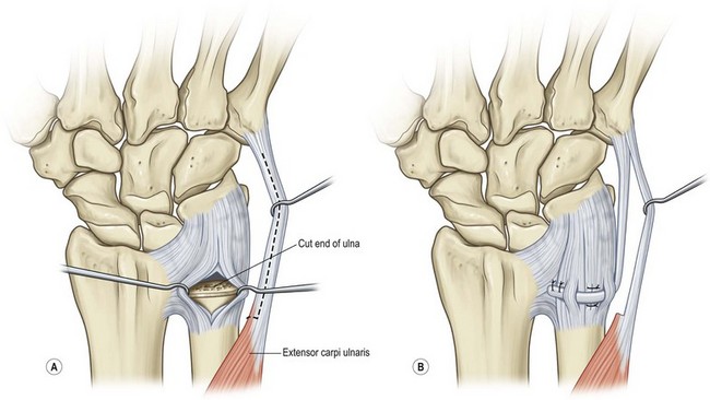
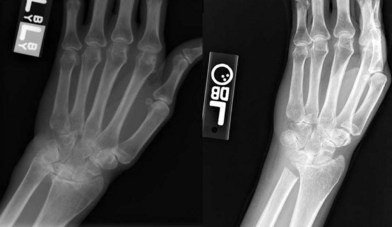
Alternatively, a distally based slip of the ECU is elevated. The DRUJ capsule and periosteal flaps are closed with non-absorbable sutures, and are imbricated tightly with the ulna reduced volarly. The slip of ECU is then woven through the capsule at the distal end of the ulna, and is then sutured to the dorsal ulnar aspect of the radius. This sling helps stabilize the ulnar stump, and also reduces carpal supination (Fig. 19.29). Pre- and postoperative radiographs are shown in Figure 19.30.
Secondary procedures
Hints and tips
Distal ulna resection
• Create a dorsal bevel after making the transverse osteotomy, and smooth with a rasp to prevent attrition ruptures
• Distal ulna resection should be performed at the time of extensor tendon transfers if the distal ulna was the source of extensor tendon rupture
• A stabilizing procedure should be performed to decrease recurrent dorsal subluxation, minimize radioulnar convergence, and reduce carpal subluxation.


Stay updated, free articles. Join our Telegram channel

Full access? Get Clinical Tree


