Cleft and craniofacial orthognathic surgery

30 Cleft and craniofacial orthognathic surgery


Introduction


Orthognathic surgery is the term used to describe surgical movement of the tooth-bearing segments of the maxilla and mandible. Candidates for orthognathic surgery have dentofacial deformities that cannot be adequately treated with orthodontic therapy alone. Children with cleft lip and palate as well as certain craniofacial anomalies are especially prone to develop malocclusion. Indeed, where approximately 2.5% of the general population have occlusal discrepancies that warrant surgical correction, 25–30% of patients who undergo surgical correction of cleft lip and palate in infancy will have severe enough midface retrusion to require orthognathic surery.1 Maxillary hypoplasia resulting in class III malocclusion is the typical deformity seen in patients with cleft and craniofacial deformities, but class II malocclusion, anterior open bites, occlusal cants and many other dentofacial deformities can also occur. Regardless of the etiology, patient examination and treatment-planning principles remain the same. The goal of orthognathic surgery, therefore, is to establish ideal dental occlusion with the jaws in a position that optimizes facial form and function.


Basic science


Growth and development


Timing of orthognathic surgery in the pediatric patient is key to good and predictable outcomes and is mediated by the development and maturation of the craniofacial skeleton. The foundation of maxillofacial growth relies on a complex interplay between genetic processes and micro- and macroenvironmental factors which must be understood to plan orthognathic procedures on patients with clefts and craniofacial disorders.


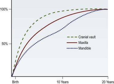
The osteogenesis of the maxillofacial skeleton occurs by way of two well-understood processes: intramembranous ossification and endochondral ossification. The cranial vault, upper face, midface, and a majority of the mandible arise from the former mechanism. Although there is a great amount of variability between individuals and genders, skeletal maturation generally progresses in a cranial-to-caudal direction with the cranial vault reaching close to adult size in early adolescence, followed closely by the upper face in the early teen years, the maxilla in the mid-teens, and the mandible in the late teen years (Fig. 30.1).3


Dental eruption patterns proceed in a similar stepwise fashion, and the transition from mixed dentition (6–12 years of age) to permanent dentition (12–20 years of age) mirrors the maturation of the maxillofacial skeleton. Indeed, midface and lower face development is, in part, mediated by the budding deciduous and permanent dentition, providing regional signals to the alveolus and stimulating bony deposition. During this period, an alteration of tooth position can, in turn, alter the direction of growth of both the maxilla and mandible. Orthodontists take advantage of this active phase of development through their use of braces, palatal expanders, and various external devices to alter maxillary and mandibular growth trajectories.4 For this reason, surgical intervention is usually delayed until skeletal maturity is reached and orthopedic movements are no longer effective.


Diagnosis/patient preparation


Preoperative evaluation


The history and physical examination


It is important to obtain a thorough medical, dental, and surgical history from every patient. Systemic diseases such as juvenile rheumatoid arthritis, diabetes, and scleroderma can affect treatment planning. With jaw asymmetries, a history of hyperplasia or hypoplasia from syndromic, traumatic, postsurgical or neoplastic etiologies affects treatment considerations. Each patient should be questioned regarding symptoms of temporomandibular joint disease or myofascial pain syndrome. Motivation and realistic expectations are important for an optimal outcome. It is likewise important for patients to have a clear understanding of the procedure, recovery, and anticipated result. In younger patients, a family discussion in terms they can understand helps to alleviate preoperative anxiety. Orthognathic surgery is a major undertaking, and the patient and family must be appropriately motivated to undergo necessary preoperative and postoperative orthodontic treatment in addition to the surgery itself.


A complete physical exam should be performed on every patient prior to surgery. The frontal facial evaluation begins with the assessment of the vertical facial thirds (richion to glabella, glabella to subnasale, and subnasale to menton) and the horizontal facial fifths (zygoma to lateral canthus, lateral to medial canthi, and intracanthal segment). The most important factor in assessing the vertical height of the maxilla is the degree of incisor showing while the patient’s lips are in repose. Males should show at least 2–3 mm, whereas as much as 5–6 mm is considered attractive in females. If the patient shows the correct degree of incisor in repose, but shows excessive gingiva in full smile, the maxilla should not be impacted. It is more important to have correct incisor show in repose than in full smile. If lip incompetence or mentalis strain is present, it is usually an indicator of vertical maxillary excess.


The inferior orbital rims, malar eminence, and piriform areas are evaluated for the degree of projection. These regions often appear deficient in cleft patients, and maxillary advancement is therefore indicated; if they are prominent, posterior repositioning may be necessary. The alar base width should also be assessed prior to surgery since orthognathic surgery may alter this width which, in turn, may accentuate any asymmetries associated with a cleft nasal deformity. Asymmetries of the maxilla and mandible should be documented on physical examination, and the degree of deviation from the facial midline noted.


The profile evaluation focuses on the projection of the forehead, malar region, the maxilla and mandible, the nose, the chin, and the neck. An experienced clinician can usually determine whether the deformity is caused by the maxilla, the mandible, or both simply by looking at the patient. This assessment is made clinically and verified at the time of cephalometric analysis. The intraoral exam should begin with an assessment of oral hygiene and periodontal health. These factors are critical for successful orthodontic treatment and surgery. Any retained deciduous teeth or unerupted adult teeth are noted. The occlusal classification is determined, and the degrees of incisor overlap and overjet are quantified. The surgeon should assess the transverse dimension of the maxilla, as prior cleft palate repair will often result in transverse growth restriction. If the mandibular third molars are present, they must be extracted 6 months prior to sagittal split osteotomy. Any missing teeth or periapical pathology should be noted, as should any signs or symptoms of temporomandibular joint dysfunction. These issues should be addressed prior to proceeding with orthognathic surgery. The term “dental compensation” is used to describe the tendency of teeth to tilt in a direction that minimizes dental malocclusion. For example, in a patient with an overbite (Angle class II malocclusion), lingual retroclination of the upper incisors and labial proclination of the lower incisors minimize the malocclusion. The opposite occurs in a patient who has dental compensation for an underbite (Angle class III malocclusion). Thus, dental compensation, which is often the result of orthodontic treatment, will mask the true degree of skeletal discrepancy. Precise analysis of the dental compensation is done on the lateral cephalometric radiographs.


If the patient desires surgical correction of the deformity, presurgical orthodondics will upright and decompensate the occlusion, thereby reversing the compensation that has occurred. This has the effect of exaggerating the malocclusion, but it also allows the surgeon to maximize skeletal movements. If the patient is ambivalent or not interested in surgery, mild cases of malocclusion may be treated by further dental compensation, which will camouflage the deformity and restore proper overjet and overbite. The importance of a commitment to surgery prior to orthodontics lies in the fact that dental movements for decompensation and compensation are in opposite directions, so this decision needs to be made prior to orthodontic therapy.5


Patient selection


Identifying the proper patient for orthognathic surgery is a key step to ensure satisfaction and successful outcomes. This includes amassing considerable data beyond a simple history and physical exam and should be coordinated with other members of the cleft/craniofacial team.


Cephalometric and dental evaluation


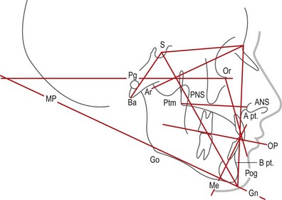
A cephalometric analysis and comparison to normative values can help the surgeon plan the degree of skeletal movement needed to achieve both an optimal occlusion and an optimal aesthetic result. A lateral cephalometric radiograph is performed under reproducible conditions so that serial images can be compared. This film is usually taken at the orthodontist’s office using a cephalostat, an apparatus specifically designed for this purpose, and a head frame to maintain consistent head position. It is important to be certain the surgeon can visualize both the bony and soft-tissue features in order to facilitate tracing every landmark. Once the normal structures are traced, several planes and angles are determined (Fig. 30.2).
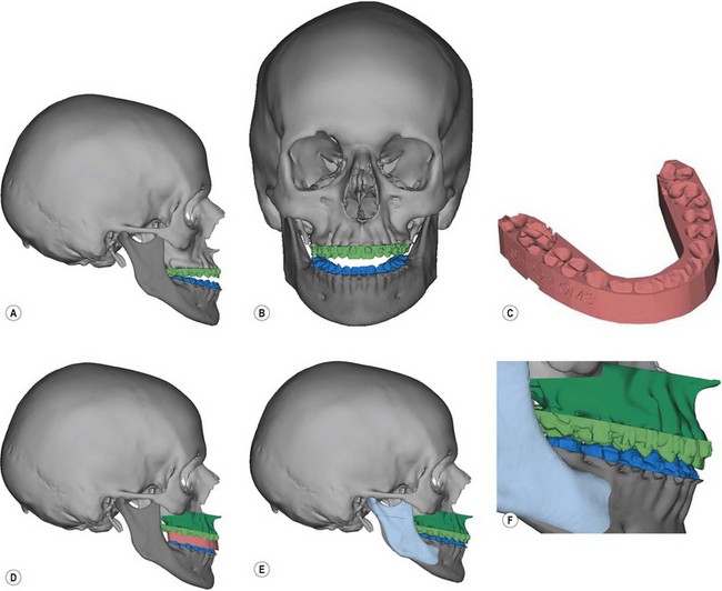


The sella–nasion–subspinale (SNA) and sell–nasion–supramentale (SNB) are the two most important angles in determining the positions of the maxilla and mandible relative to each other as well as the cranial base. These angles are determined by drawing lines from sella to nasion to A point or B point, respectively. By forming an angle with the sella and nasion, this position is referenced to the cranial base. A point provides information about the anteroposterior position of the maxilla. If the SNA angle is excessive, the maxilla exhibits an abnormal anterior position relative to the cranium. If SNA is less than normal, the maxilla is posteriorly positioned relative to the cranial base. The same principle applies to the mandible: B point is used to relate the mandibular position to the cranial base. The importance of the cranial base as a reference is that it allows the clinician to determine if one or both jaws contribute to a noted deformity. For example, a patient’s class III malocclusion (underbite) could develop from several different etiologies: a retrognathic maxilla and a normal mandible as is common in cleft patients, a normal maxilla and a prognathic mandible, a retrognathic mandible and a more severly retrognathic maxilla, or a prognathic maxilla and a more severely prognathic mandible. All of these conditions yield a class III malocclusion, yet each requires a different treatment approach. The surgeon can delineate the true etiology of the deformity by the fact that the maxilla and mandible can be independently related to a stable reference, the cranial base. Next, cephalometric tracings are performed.


Cephalometric tracings give the surgeon an idea of how skeletal movements will affect one another as well as the soft-tissue profile. They also allow the surgeon to determine the distances the bones will be moved to achieve the goals of specific procedure. Different tracing methods using acetate paper are used for isolated maxillary, isolated mandibular, or two-jaw surgeries. Much of the traditional hand cephalometric tracing, however. has given way to computer-aided cephalometric analysis which allows the surgeon to position the maxilla and mandible electronically on the cephalogram while recording the soft-tissue changes and measuring the degree of repositioning.


Complete dental records, including mounted dental casts, are needed to execute preoperative model surgery and fabricate surgical splints. Casts allow the surgeon to evaluate the occlusion both before and after articulation into proper positions. Analysis of new occlusion gives the clinician an idea of how intensive the presurgical orthodontic treatment plan will be. Casts also allow the clinician to distinguish between absolute and relative transverse maxillary deficiency. Absolute transverse maxillary deficiency presents as a posterior crossbite with the jaws in class I relationship. A relative maxillary transverse deficiency is commonly seen in a patient with a class III malocclusion. A posterior crossbite is observed in this type of patient, raising suspicions of inadequate maxillary width. However, as the maxilla is advanced or the mandible retruded, the crossbite is eliminated. Articulation of the casts into a class I occlusion allows the surgeon to distinguish easily between relative and absolute maxillary constriction.


Model surgery


Using the cephalometric tracings as a guide, the next step is to reproduce the maxillary and/or mandibular movements on articulated dental models. This allows for the fabrication of occlusal splints to be used intraoperatively to guide jaw repositioning in preparation for osteosynthesis. Model surgery begins by obtaining accurate casts of the patient’s occlusion. If the surgeon does not have a dental laboratory, the orthodontist will obtain the casts. The success of the technical portion of orthognathic surgery correlates directly with the accuracy of the model surgery and splint fabrication.


Isolated maxillary and two-jaw surgery


A face bow is a device used to relate the maxillary model accurately to the cranium on an articulator. If a maxillary osteotomy is being performed, one set of models should be mounted on an articulator using the face bow. Two other sets of models are used in treatment planning. Next, an Erickson model block is used to measure the current position of the maxillary central incisors, cuspids, and the mesiobuccal cusp of the first molar. The face bow-mounted maxillary cast is placed on the model block. The maxillary model is then measured to the tenth of a millimeter vertically, anteroposteriorly, and end-on. By having numerical records in three dimensions, the surgeon can reproduce the maxillary cast’s exact location, as well as determine a new location. Reference lines are circumferentially inscribed every 5 mm around the maxillary cast mounting. The distances the maxilla will move in an anteroposterior, lateral, and vertical direction have been determined from the previous cephalometric exam. These numbers are added or subtracted from the current values measured on the model block to determine the new three-dimensional position of the maxillary cast. The occlusal portion of the maxillary cast is removed from its base using a saw. As much plaster is removed from the cast as is necessary to accommodate the new position of the maxilla. Once the model block verifies the maxilla is in its new position, the cast is secured with sticky wax or plaster to the mounting ring. Now it can be placed on the articulator. At this point, the surgeon has a mounting of the postoperative maxilla related to the preoperative mandible. An acrylic splint is made at this point. This splint is called the intermediate splint and is used in the operating room to index the new position of the maxilla to the preoperative position of the mandible. A second mounting with the casts in the occlusion desired by the orthodontist is used to make a final splint that represents the new position of the mandible to the repositioned maxilla. This is fabricated in a manner similar to the splint for isolated mandibular surgery. If the occlusion is good, intercuspal position can be used to position the mandible without the splint.


3D CT modeling


There are several computer-assisted design (CAD) programs that are now commercially available that can assist the surgeon with some or all of the preoperative patient preparation. A computed tomography (CT) scan is obtained with the patient wearing a bite jig that correlates natural head position to the three-dimensional (3D) CT image of the patient’s face. Although conventional helical CT scans with fine cuts through the face are ideal, cone beam CT scans offer a comparable image quality with considerably less cost and radiation exposure (50 µSv compared to 2000 µSv). A cephalometric analysis can then be performed as well as simulated movements of the jaws and chin in any dimension. Once the osteotomy movements are verified by the surgeon, CAD/CAM technology is used to fabricate surgical splints for the patient. If necessary, 3D models of the patient can be made showing the exact proposed movement (Fig. 30.3). Some systems can actually “wrap” a 2D digital image around the soft-tissue envelope of the 3D CT image, thus replicating a 3D image of the patient’s face in color.


In these authors’ experience, 3D CT modeling has demonstrated improved accuracy in diagnosis and treatment. The elimination of traditional model surgery saves the surgeon time in patient preparation. Finally, the 3D aspect of this treatment-planning approach enhances the surgeon’s ability to predict how osteotomies may affect soft tissue of the face. These advantages facilitate the ability of the plastic surgeon to provide optimal care for these patients.


Developing a treatment plan


Once the data are obtained, the surgeon can determine which abnormalities the patient exhibits and the extent to which these features deviate from the norm. The treatment plan is the application of these data to provide the best aesthetic result while establishing a class I occlusion. The goal is not to “treat the numbers” in an attempt to normalize every patient. The appearance of the soft-tissue envelope surrounding the facial skeleton is the most crucial factor in determining the aesthetic success of orthognathic procedures, and the jaws should be positioned so they provide optimal soft-tissue support.


Historically, skeletal movements that expanded the soft tissue of the face were less stable, so posterior and superior movements were preferred. Although these movements were more stable, they resulted in contraction of the facial skeleton with the associated soft-tissue features of premature aging. Since the introduction of rigid fixation systems, osteotomies that result in skeletal expansion have been achieved with a great degree of predictability. An attempt is made to develop a treatment plan that will expand or maintain the preoperative volume of the face. If a superior or posterior (contraction) movement of one of the jaws is planned, an attempt should be made to neutralize the skeletal contraction with an advancement or inferior movement of the other jaw or the chin. It is important to avoid a net contraction of the facial skeleton as this may result in a prematurely aged appearance.

Stay updated, free articles. Join our Telegram channel

Feb 21, 2016 | Posted by in General Surgery | Comments Off on Cleft and craniofacial orthognathic surgery

Full access? Get Clinical Tree

Get Clinical Tree app for offline access