Fig. 49.1
Sheets of atypical melanocytes with abundant eosinophilic cytoplasm comprise rhabdoid melanoma
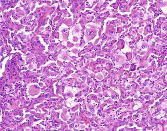
Fig. 49.2
The tumor cells have marked nuclear atypia and abundant densely eosinophilic cytoplasm. The appearance is somewhat reminiscent of rhabdomyoblasts
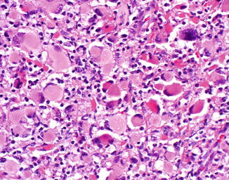
Fig. 49.3
The tumor cells have marked nuclear atypia and abundant densely eosinophilic cytoplasm. The appearance is somewhat reminiscent of rhabdomyoblasts

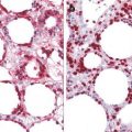
Fig. 49.4
The presence of perinuclear eosinophilic hyaline globules (arrow) which peripherally displace the nucleus is a characteristic feature
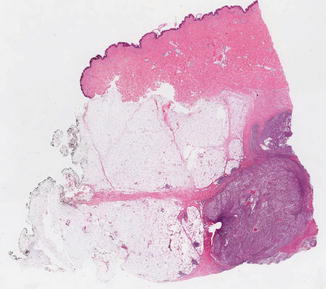
Fig. 49.5


Metastatic rhabdoid melanoma displays a nodular proliferation of melanocytes in the subcutis similar to cutaneous metastases of conventional melanoma
Related posts:


Stay updated, free articles. Join our Telegram channel
Full access? Get Clinical Tree
