Fig. 38.1
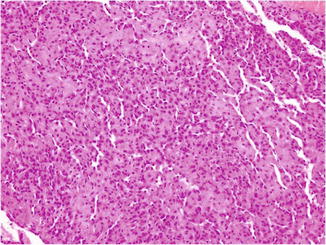
Retiform hemangioendothelioma. A vascular tumor composed of elongated arborizing blood vessels involving the dermis and subcutis

Fig. 38.2
Retiform hemangioendothelioma. The arborizing blood vessels are arranged in a pattern reminiscent of the normal rete testis architecture

Fig. 38.3
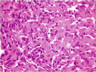
Retiform hemangioendothelioma. The arborizing blood vessels are lined by monomorphic endothelial cells with prominent protuberant nuclei having a characteristic hobnail-like appearance

Fig. 38.4
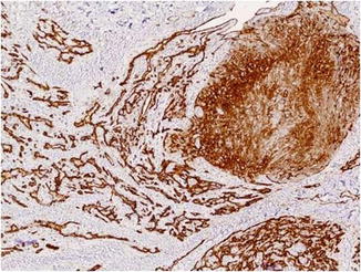
A vascular tumor with mixed features of Dabska’s tumor and retiform hemangioendothelioma showing positivity with CD31
Immunohistochemically, the tumor cells react with endothelial markers such as CD31 (Fig. 38.4), CD34, factor VIII-related antigen, and ERG. Staining for CD34 is usually stronger than that for other vascular markers. Claudin-5, a tight-junction protein, has recently been proposed as a reliable vascular marker. Cytokeratins and smooth muscle actin are negative. Most lymphatic markers, including podoplanin (D2-40) and VEGFR-3, are negative. In one case report, HHV8 DNA sequences were claimed to be detected, but in general experience, RH are negative for HHV8.
Differential Diagnosis
The diagnosis is only a histological one. The differential diagnosis prior to biopsy includes lymphoma, dermatofibrosarcoma protuberans, hemangioma, bacillary angiomatosis, cutaneous metastases, blue-rubber bleb nevus syndrome, Kaposi sarcoma, targetoid hemosiderotic hemangioma (hobnail hemangioma), malignant endovascular papillary angioendothelioma (Dabska’s tumor), and cutaneous angiosarcoma. In cases with overlapping features with cutaneous angiosarcoma, the diagnosis is primarily based on the degree of nuclear atypia, the number of mitosis, and the layering of endothelial cells.
Stay updated, free articles. Join our Telegram channel

Full access? Get Clinical Tree


