The authors deal with different kinds of patients exhibiting different anatomic arrangements of the structures in their neck, different ages, and different expectations; there are several techniques from which to choose. Patients have no idea of the deformity they show. They demand a fast recovery with the best possible result that is long lasting. Because of the diversity of the involved elements, a neck lift is an extremely complex challenge for the surgeon, demanding an accurate technique and common sense.
Key points
- •
To restore neck contour, we do not favor the following procedures: Submandibular gland resections: Operations on the submandibular gland may yield severe complications, and the benefits are not worth the risk. Digastric muscle operations: We see no advantages to operations on the digastric muscles. They are masticatory muscles.
- •
To provide the best outcome for the patient, the surgeon must respect the complexity and variance of neck defects, the limitations of each case, and the surgeon’s own limitations.
Editor Commentary: Claudio had the opportunity to dissect dozens of cadavers and as a result of this important study, he described the three typical anatomic appearances and the associated decussation of the platysma muscles. In this chapter he takes us through his personal evolution in necklifting based upon the clinical findings and working towards a desirable clinical outcome. Even Claudio’s relatively simple maneuver of adding 1cm to the submental incision has been helpful in allowing improved vision to the relevant submental anatomy with no consequence in patient satisfaction.
A neck deformity is the main complaint of patients who seek a rhytidoplasty. Patients present with diverse ages, different deformities, and variable aspirations, which brings a tough challenge for the surgeon.
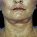
Observing Fig. 1 , the differences among patients are noticeable, showing variable deformities, such as skin excess, fat amount and distribution, bone structure, and so forth. Therefore, it is mandatory to provide a very personal surgical plan for each one.
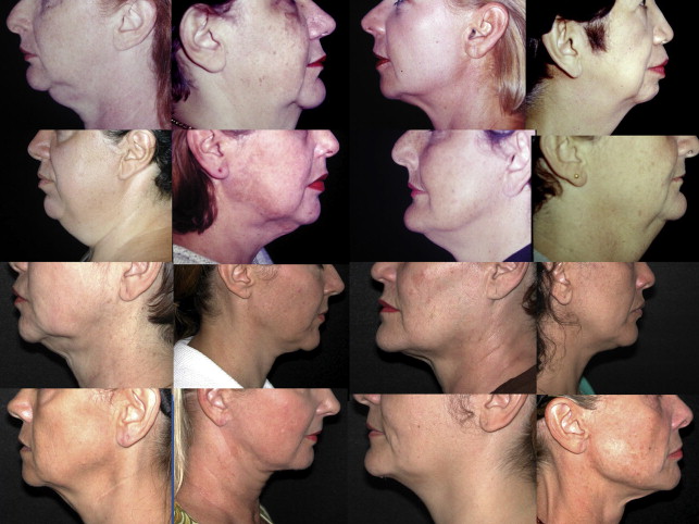
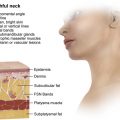
Skin excess together with fat and muscle superficial muscular aponeurotic system (SMAS)/platysma alterations are responsible for neck contour deformities. These alterations will be more or less noticeable according to the bone structure, skin quality, position of hyoid bone, and the submandibular gland. According to the magnitude of these factors, the surgical plan is aimed at the elimination/attenuation of the deformities. To plan the procedure appropriately, one must respect the limitations of each case and even the surgeon’s limitations.
We do not favor submandibular gland resection and/or digastric muscle operations. The operations on the submandibular gland may yield severe complications, and the benefits are not worth the risk. We do not see the advantages of operations on the digastric muscles. They are masticatory muscles.
In most of the cases, we unite the skin dissection at the suprahyoid region.
We open the submental area whenever there is alteration at the median line. In doubt, we always open it. We never regretted opening the neck; in some cases, we would have had a better result if we had opened the neck. It is important to state that we never had a complaint on the imperceptible scar below the chin.
When there is severe deformity in the jaw line, we recommend an ample SMAS dissection.
When lipectomies are performed, they are done so in a limited fashion in order to avoid skin retractions or artificial results and skin undulations.
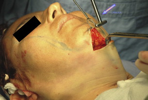
For submental aspiration, we use Pontes cannula ( Fig. 1 ) that, in our opinion, facilitates the fat removal evenly, in an open manner. We do not favor subplatysmal defatting because there is a natural space between the platysma muscle and the mylohyoid muscle. If fat is removed, the muscles will heal and a deformity will occur that is very difficult to correct.
When fat is removed, the medial fibers of the platysma muscles are dissected to evaluate the thickness, anatomic behavior, and flaccidity of the muscles. A lateral dissection is done, and the muscle excess is carefully observed and removed. A medial suture is performed following a medial section as described by Aston using scissors as proposed by Aboudib. For us, it is extremely important to remove the muscle in excess because the approximation of the medial fibers in a great percentage of patients with muscle excess will show redundancy of the muscle if it is not reduced ( Fig. 2 ). That is why there is much importance to knowing and understanding the anatomic knowledge of the anatomy of the medial fibers of the platysma muscle.
Some remarks on the anatomy of medial fibers of platysma muscle
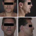
In 1980, we published an article on platysma anatomy where we dissected 100 formalized cadavers. The initial purpose of the study was to observe the mandibular ramus of the facial nerve. When we were studying the photographs, we noticed different types of distribution of the medial fibers of the platysma muscle in the midline. We noticed the fibers were joined or separated. Then we classified the fibers into 3 different groups.
- 1.
Type I: The fibers interlace 1 to 2 cm below the chin ( Fig. 3 A ).
Fig. 3
( A ) Platysma type I fibers decussate about 3 cm below the chin being more separated or less separated at the suprahyoid region. ( B ) Platysma type II fibers are joined at the submental region behaving as a single muscle. ( C ) Fibers do not interlace at the suprahyoid region. The muscular fibers may be more or less separated presenting different grades of thickness.Related posts:
 Nonexcisional, Minimally Invasive Rejuvenation of the Neck
 Progressive Tunnelizations in Neck Face Lift Detachment
Lore’s Fascia a Strong Fixation Point for Neck Rejuvenation Procedures
Progressive Tunnelizations in Neck Face Lift Detachment
Lore’s Fascia a Strong Fixation Point for Neck Rejuvenation Procedures
 Managing the Components of the Aging Neck
 Multidimensional Evaluation and Surgical Approaches to Neck Rejuvenation
 Neck Lift Technique

Stay updated, free articles. Join our Telegram channel

Full access? Get Clinical Tree


