Reconstructive surgery of the mutilated hand

12 Reconstructive surgery of the mutilated hand


Introduction


Management of the mangled upper extremity is complex, demands special skills and expertise, and is facilitated by a team approach with participation of all those dedicated to upper extremity surgery. When adequate facilities, equipment, and surgical expertise are not available to manage the patient’s degree of injury, the patient should be transferred with the extremity splinted. If the limb is ischemic, or if part of the limb is amputated, the extremity should be cooled. The ischemic limb should be carefully covered in ice, with a protective barrier to prevent frostbite injury. The amputated part should be wrapped in a saline-soaked gauze, placed on ice, and sent with the patient. The ideal temperature is +4°C. Dry ice or alcohol should not be used to aid in cooling during transport of the part as this can cause actual freezing of the tissues.


Resuscitation of the patient is a priority over addressing the extremity injury. However, in the event of a coexisting life-threatening injury, the extremity injury must not be neglected. Assessment of vascularity, realignment, and splinting are not time-consuming and should be done as soon as possible. Provisional revascularization through shunting may be an option to restore circulation to the extremity quickly while the trauma team simultaneously addresses the coexisting injuries. Consent of the patient should initially be obtained for wide debridement of the wound; internal versus external fixation; repair or reconstruction as indicated of nerves, vessels, tendons, and muscles; use of vein grafts for arterial and venous reconstruction; possible donor sites for flap coverage; and primary amputation. The selected treatment is based on the intraoperative findings.


The initial treatment includes meticulous debridement, wound irrigation, wound culture, antibiotic administration, and tetanus prophylaxis. Copious irrigation of the wound coupled with debridement is important because it removes foreign bodies and decreases the bacterial load. High-pressure and/or pulsed lavage of the soft tissues is to be avoided in these types of injuries, as it may force debris and bacteria into the tissues and has a tendency to force fluid into the tissues, potentially creating more edema in the postoperative period. The cultures most predictive of later infecting organisms are the postirrigation cultures. Antibiotics will help reduce infection and should be administered in the emergency department and continued intravenously for at least 72 hours. Antibiotic coverage should be effective against both Gram-positive and Gram-negative organisms. The authors currently prefer to use a first-generation cephalosporin and an aminoglycoside in combination. In cases of soil contamination, as in farm injuries, coverage against anaerobic organisms should always be added. High-dose penicillin provides excellent anaerobic coverage. The possibility of gas gangrene should also be considered in these cases. The antibiotic regimen should subsequently be modified depending on the culture results. Tetanus prophylaxis should be administered in accordance with the status of previous immunization.


Debridement


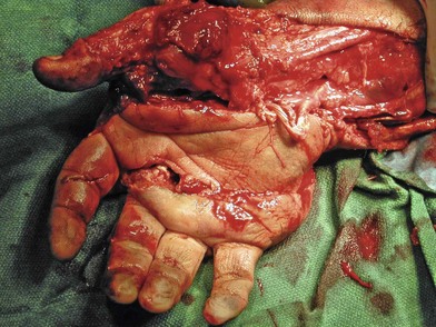
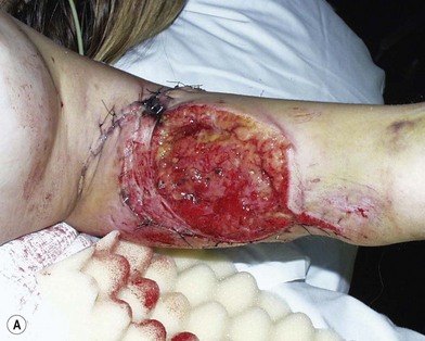
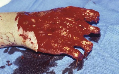
Radical debridement with elimination of all nonviable tissue is the crucial step in the management of these injuries. This is performed under tourniquet control to provide the best visualization of the extent of injury and to prevent iatrogenic injury to intact structures. Once nerves and vessels are identified, the tourniquet may be released to assess the viability of the remaining tissue better (Fig. 12.1).


Skin and subcutaneous tissues are sharply debrided to bleeding edges. Muscle is debrided until bright red blood is seen. Color and twitch are used to assess muscle tissue; however, these characteristics are not totally reliable in determining viability. If the muscle does not bleed, it is dead and should be debrided. Small bone fragments devoid of soft-tissue attachments are avascular and should be discarded unless they constitute part of an articular surface. Contused or contaminated nerves are left in continuity. Superficial dissection above the epineurium can be carefully done to remove foreign material if it is present. Cut nerves are debrided to healthy-appearing fascicles. At the time of reconstruction, this is done under microscopic visualization because a primary repair or nerve graft will fail if the entirety of the injured segment is not resected.


image Video1


The wound is copiously irrigated. Tissue margins are re-evaluated, and further debridement is performed as necessary. Hemostasis should be done cautiously. Do not cauterize vessels that may be needed for arterial or venous reconstruction. Side branches may be controlled with small sutures or vascular clips. Arterial debridement is performed for cut or thrombosed vessels. Definitive debridement of arteries that are to be reconstructed is done with the tourniquet released. At the completion of debridement, assess and record what structures remain intact and what the functional losses are. Some of the remaining intact muscles may be suitable for immediate tendon transfer to replace important functional losses.


Provisional revascularization


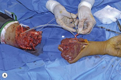
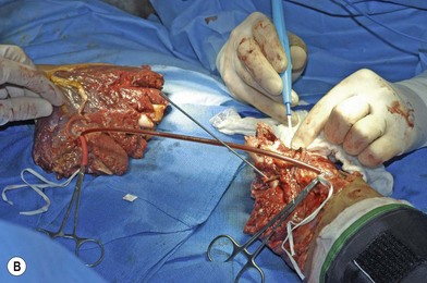
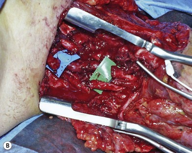
When the extremity is amputated or ischemic and the delay to surgery or anticipated length of time for debridement and definitive skeletal stabilization exceeds 6 hours, provisional revascularization can be done with use of a shunt, such as a Javid, Ishihara, or comparable segment of plastic tubing (Fig. 12.2). While different shunts are available, their use is similar. Whichever shunt is chosen (primarily by what might be available for use by the vascular surgeons), it is generally flushed with heparinized saline and then clamped. The concentration of this is usually 100 units heparin per cc saline. The shunt is then carefully slipped into the ends of the vessels to be shunted (artery to artery). The shunt is either held in place with moist umbilical tapes placed around the vessels and clamped tight with a piece of red rubber catheter (Fig. 12.2), or some shunts come with a special clamp which goes around the artery and holds it flush on the shunt. While generally utilized for only short periods of time (i.e., minutes to a few hours), these types of shunt have recently been utilized to great effect in the conflict in the Middle East for up to 8 hours on major arteries, without systemic heparinization. In this application, however, their use should only be necessary for the time it will take to perform an initial debridement and bony fixation. Another option is rapidly to perform reversed vein grafting between the arterial ends to re-establish arterial inflow. After definitive skeletal stabilization, the length of the vein graft can be adjusted and the anastomosis revised.


Skeletal stabilization


Definitive stable internal fixation should be done immediately when possible to allow therapeutic access to the extremity for wound care and early joint range of motion when applicable. In general, the use of external fixation is to be avoided. These devices carry the risk of neurovascular injury during pin placement, restrict circumferential access to the extremity, and may limit rehabilitation. They may be useful as a temporary solution for severely contaminated wounds with extensive soft-tissue injury, however. External fixation should not be used for the reason of avoiding exposed hardware. Potentially exposed hardware should be addressed with appropriate soft-tissue coverage.


Diaphyseal fractures of the arm and forearm are usually amenable to plate fixation. If the fracture is segmental or comminuted over a large segment, a flexible interlocking intramedullary nail can be used. Fractures should be reduced anatomically with respect to rotational alignment. One must carefully check pronation and supination prior to final fixation to avoid placing the hand in a poor position. Comminuted bone fragments without soft-tissue attachments should be removed. Shortening of the humerus up to 5 cm and of the radius and ulna up to 4 cm is acceptable. Indications for shortening include significant comminution over a segment with bone and soft-tissue loss. Bony shortening has the advantage of allowing primary repair of vessels and, more importantly, of nerves.


Complex diaphyseal fractures include Galeazzi fractures (radial diaphyseal fracture with disruption of the distal radioulnar joint), Monteggia fractures (proximal ulna fractures with radial head dislocation or radial head or neck fracture), and Essex–Lopresti injuries (radial head fracture with associated rupture of the interosseous membrane to the level of the wrist). These fractures must be restored to anatomic length to restore joint congruity and alignment.


Diaphyseal fractures of the hand are fixed with Kirschner wires, with attention to maintaining rotational alignment. Shortening of the metacarpals or phalanges between 5 and 6 mm may be necessary in replantation. For other injuries, length can be restored or maintained with internal or external fixation and primary or delayed bone grafting. In general, initial use of plate fixation is not recommended in the hand, particularly in the phalanges. This requires more stripping of the periosteum and later has an adverse effect on functional outcome because of potential tendon adhesions around the plate.


In contrast to lower-extremity injuries, primary bone grafting is acceptable and recommended in the upper extremity. Primary iliac crest bone graft or bone allograft may be used for defects up to 4 cm. For larger defects, vascularized bone grafts, in particular a vascularized segment of free fibula, should be considered. With massive bone loss, creation of a one-bone forearm can be considered as a salvage procedure when other options are not available. The results of this procedure in trauma are inferior to those in limb salvage surgeries for tumor reconstruction.


Intra-articular fractures should be anatomically reduced. Stable fixation is imperative to allow early range of motion. Fixation can be done with plates, screws, K-wires, or tension band wiring as dictated by the fracture pattern and location. Articular fragments free of soft-tissue attachments should be preserved to reconstruct the joint surface. Primary bone grafting should be done to address bone defects, to enhance the stability of the fixation, and to promote bone healing. When the joint surface cannot be reconstructed owing to severe comminution or bone loss, other reconstructive options are available. At the shoulder and elbow, a primary allograft or a prosthesis can be used. At the wrist joint, primary fusion should be the first treatment option. If wrist motion is critical to the patient, vascularized free fibula, retaining the proximal articular cartilage, can be used to reconstruct the joint surface. Prosthetic devices for total wrist arthroplasty are poor at present, and historically do not do well in posttraumatic wrists regardless.


In summary, the goal of skeletal stabilization is to achieve stable anatomic fixation to allow early range of motion and rehabilitation. Soft-tissue stripping of bone is limited to only that which is necessary for fixation. Comminuted fragments with attached soft tissue are retained. Bone defects are treated primarily whenever possible. The potential of exposed hardware should not limit the choice of fixation. Exposed bone, hardware, or joints are addressed with appropriate soft-tissue coverage, whether it be pedicled or free.


Vascular reconstruction


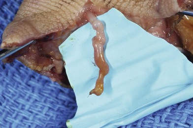
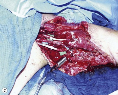
Definitive revascularization is done once skeletal stabilization is completed. Lacerated vessels are resected to healthy-appearing vessel wall both proximally and distally. Contusion along the adventitia suggests injury within the intimal layer as well. A “ribbon sign” (convoluted or tortuous course of the digital vessels) indicates injury to the media layer of the vessel and requires resection of the length of the involved area and reversed vein grafting (Fig. 12.3). Inflow is assessed, and if it is not adequate, the vessel should be resected more proximally until pulsatile flow is achieved.


When possible, primary repair is preferred. However, it is better to resect appropriately and to use a reversed vein graft than to perform a primary repair under tension. Reversed vein grafts are available from several sites. The most commonly used for long segments in the upper extremity is the saphenous vein. For reconstruction in the hand, local grafts from the dorsal or volar forearm can be used.


Reconstruction of the superficial palmar arch and its multiple common digital arteries may be difficult. Branched vein grafts from the dorsum of the foot and use of the subscapular artery and its branches have been described for this. The descending branch of the lateral femoral circumflex artery may also be utilized as a smaller arterial graft to make use of its branches to reconstruct the arch. The conventional method is to harvest two Y vein segments from the volar forearm and to perform end-to-end or end-to-side anastomoses as necessary. Vein grafts should be routinely used to reconstruct venous outflow if primary repair cannot be achieved. There is a minimal role for artificial grafts for vascular reconstruction in the upper extremity distal to the axillary artery, particularly in contaminated wounds.


When the need for coverage and vascular repair present themselves simultaneously, one can consider “flow-through” free flaps both to bridge the arterial defect and to obtain coverage at the same time. The radial forearm free flap is very useful in this regard, as the radial artery offers an excellent choice for a long bypass.1 Due to trauma to the forearm, however, this may not be an appropriate choice of flap. In this case, the anterolateral thigh flap with its long segment of the descending branch of the lateral femoral circumflex can be utilized.2,3 Small venous free flaps with arterialized segments of vein can be utilized to great utility for revascularization of fingers in the face of a soft-tissue loss volarly.4,5


Musculotendinous reconstruction


Primary tendon repair is preferred when permitted by the injury. There is often a crush or avulsion component to the injury which may preclude primary repair. In these cases, treatment options include primary or delayed reconstruction with tendon grafts or Silastic tendon rods. Immediate reconstruction is favored with the use of available donor tendons (palmaris, plantaris, local tendons that cannot be reconstructed, and toe extensors).


In flexor tendon injuries in the hand, priority is usually given to reconstruction of the profundus tendons. If there is trauma to the distal interphalangeal joint requiring fusion, priority is rather given to the superficialis tendon. The lesser-priority tendon can be used for reconstruction of the other when necessary. It can be used as a donor tendon for other sites or for pulley reconstruction as necessary. One should pay close attention to the status of the pulleys in the fingers, and the A2 and A4 should be reconstructed with portions of tendon grafts when necessary.


Functional reconstruction of muscle deficits and tendon injuries should be done immediately whenever possible. Unrecoverable muscle function can be treated with tendon transfer, and it is preferable to perform this as a primary procedure. However, this can also be performed at a later stage of reconstruction. Delayed transfers are generally more difficult because they have to be performed through a scarred tissue bed, and this requires additional surgical procedures and further delays the patient’s rehabilitation and recovery. When no donor tendons are available for transfer, muscle function can be restored with functional free muscle transfer. The gracilis is the most commonly used muscle for functional reconstruction and can actually provide both coverage and restoration of muscle function to the fingers. Again, this can be done either primarily or at a later stage of reconstruction; however primary reconstruction is favored as available nerves and vessels are easier to locate and one is not operating through a scarred bed of tissue. This procedure will be discussed in more detail below and in Chapter 35.


Nerves


Nerves may have internal derangement without loss of anatomic continuity and they can be partially or completely disrupted. Severe contusion to the nerve with internal hemorrhage without actual division may mitigate against functional return; however contused or attenuated nerves are usually left intact. If lacerated, the nerve ends should be debrided serially under magnification until healthy-appearing fascicles appear. The resection should not be compromised in order to preserve length, as nerve regeneration will not occur through scarred nerve tissue, and nerves repaired under tension also do not regenerate well.


Mobilizing the proximal and distal stumps to achieve primary repair is not recommended because this results in devascularization of large segments of the nerve. Mobilization can be done over a 1–2-cm distance to allow repair, but to avoid repair under tension, nerve grafting is preferable. If a staged repair is planned, nerve ends are tagged with 6-0 polypropylene suture for later identification. Primary nerve grafting is recommended, however, as the orientation is much clearer at the time of injury rather than later and dissecting a nerve out of a scarred bed is technically challenging (Fig. 12.4). Common donor nerves include the sural or saphenous nerves, sensory branch of the radial nerve (if lacerated from the injury), medial or lateral antebrachial cutaneous nerves, and the posterior interosseous nerve. In multiple nerve injuries, primary nerve transfers can be performed.6 These include transfer of the anterior interosseous nerve (distal to the branch to the flexor pollicis longus) to the motor branch of the ulnar nerve and transfer of the sensory branch of the radial nerve to the digital nerves of the thumb and index finger.7 When a nerve defect is greater than 15 cm, an end-to-side neurorrhaphy of the distal stump of the injured nerve to a neighboring intact major nerve can be done, although functional outcomes of this procedure are generally poor.


Skin and soft-tissue reconstruction


Appropriate soft-tissue coverage is of paramount importance for coverage of bone, joint, tendons, neurovascular structures, and hardware. The selection of coverage should provide a gliding surface for mobile structures and enhance vascularity in the injured area.


Extensive skin and soft-tissue loss is one of the major problems in the mangled upper extremity. Isolated skin loss may be managed by split-thickness skin grafting (if not over a bony prominence, hardware, or exposed tendons). Local random skin flaps have limited utility even in superficial soft-tissue defects owing to their restricted mobility, precarious blood supply, and frequent involvement in the injury.


Soft-tissue loss can be managed in a variety of ways. A simple approach is to leave the wound open and let it heal by secondary intention with granulation tissue. However, exposed tissues sensitive to desiccation, such as nerves and tendons, will become necrotic, scarring will be promoted, and function will be compromised. Local muscle flaps are usually not suitable because of their proximity to the zone of injury, the limited amount of coverage that they provide, and the resulting functional deficit. Two-stage distant pedicled flap procedures are avoided when possible; immobilization of the reconstructed area will lead to stiffness and swelling. If this means of coverage is required, the most common sources are the pedicled groin flap, cross-arm flap, thoracoacromial flap, and abdominal flap. In mangling injuries, the need for extensive coverage, reliable vascularity of the flap, and early mobilization usually necessitates use of axial flaps (local or regional), one-stage distant pedicled flaps, or free flaps. The type of coverage depends on the site and extent of the defect. This is particularly important for bone, tendon, nerve, and hardware coverage, as well as for facilitation of future reconstructive procedures in planning a staged reconstruction. As discussed above, one should think one or two steps ahead in terms of what reconstructive procedures are potentially going to be performed later under or though the chosen flap.


Fasciocutaneous flaps or cutaneous flaps with subcutaneous tissue are recommended when tendons are exposed as this tissue facilitates tendon gliding. However, many fasciocutaneous flaps over time do not have a good cosmetic appearance (color match can be poor and they can be too bulky). The primary fasciocutaneous flaps used with acceptable cosmetic appearance include the radial forearm flap, as either a free or rotational flap; the lateral arm flap; and the groin flap. These can all be utilized as free flaps, but the groin flap is generally utilized today as a pedicled flap due to the small size and short available vascular pedicle. An alternative to the fasciocutaneous flap is a fascial flap. Donors for this include the temporoparietal fascia, the parascapular fascia, and the radial forearm fascia. The free fascial flap is placed on the defect and then covered with an unmeshed split-thickness skin graft. It has been our experience, though, that it can be difficult to elevate a fascial flap later for tenolysis or other procedures.


Muscle flaps are used when a moderate to large soft-tissue defect is present. Although muscle flaps may be harvested with a skin component, harvesting of muscle alone is preferred. This is then covered with a split-thickness skin graft. In the arm and elbow, the latissimus dorsi can be used as a one-stage distal pedicled rotational flap. Most other reconstructions are done with a free muscle transfer using the gracilis, rectus abdominis, latissimus dorsi, or serratus anterior. When a functional deficit is present, consideration should be given to use of a functional free muscle transfer to provide both functional restoration and soft-tissue coverage. The gracilis is most commonly used for this transfer in the forearm, as noted above.


Postoperative management


In the immediate postoperative period, the extremity is splinted in an appropriate position to prevent capsuloligamentous shortening and tension on repaired structures. Elevation is necessary to reduce edema and help control pain. The patient’s pain and anxiety should be adequately controlled. The ambient temperature should be at least 25°C and adjusted to the patient’s body temperature. Hydration should be sufficient to maintain urine output between 80 and 100 mL/h. Anticoagulant therapy is used by many surgeons. However, the authors are selective in using heparin; aspirin is used daily for 4–6 days after surgery. Appropriate antibiotic therapy is continued in the postoperative period and later modified as necessary according to culture results.


Extremity and flap monitoring is critical. Early recognition of arterial hypoperfusion or venous congestion will occur only in a well-monitored environment with trained nursing staff. If there is a question about tissue viability, the dressings are released and the entirety of the revascularized tissue is exposed for further assessment of perfusion, congestion, temperature, turgor, and color with Doppler examination. Release of the dressings alone may be sufficient to alter the vascular issue. If viability is still in question after 30 minutes, immediate re-exploration and further assessment may prevent failure of the revascularized tissue, whether it be the extremity or a free flap.


Early and motivated rehabilitation of the extremity is an important factor in achieving a successful outcome. Early motion reduces edema, adhesions, and scarring. It prevents muscle atrophy and facilitates healing of the soft tissues by remodeling of collagen fibers. The details of the rehabilitation program are determined by the existing injuries and the reconstruction procedure.


Basic science/disease process


Upper extremity function and facial appearance and expression are the two central defining features of our humanity. Movement of the hand through space along with precise grasp, pinch, and positioning allows us to perform the most rudimentary to the most highly complex tasks to actualize the entire range of our wishes, dreams, and ambitions. Man without hands is akin to a plane without wings. Nearly all tasks combine positioning of the hand with shoulder, elbow, and wrist motion followed by intricate manipulation of finger joint position with both intrinsic and extrinsic muscle force modulation.


Injuries to any part of the upper extremity, no matter how minor, may immediately compromise and chronically debilitate the user’s ability to perform. Witness the enormously negative impact of work-related injuries on our economy. Productivity is reduced, medical costs are escalated, and the work environment is disrupted, not to mention the enormous personal and family suffering caused by pain and loss of function. Some have estimated the cost to our economy from upper extremity trauma to run in the hundreds of billions of dollars. In light of the significantly problematic consequences that result from extensive upper extremity injuries, it is paramount that early diagnosis and, more important, strategically sound treatment be instituted from the outset.


Extensive skin and soft-tissue trauma along with composite injuries involving multiple structures must be managed actively, there being few circumstances in the hand in which open wound management and healing by secondary intention play a role. Maximum preservation of motion and sensibility should be ever present in the mind of the reconstructive surgeon from the moment of first evaluation, with prompt wound closure given high priority.


The term “mangled” is commonly used to describe the hand and upper extremity after major trauma. Gregory et al. used the term “mangled” to describe a severe injury to at least three of the four organ and tissue systems of skin, bone, vessel, and nerve.8 According to the Oxford English Dictionary, to mangle is “to reduce by cutting, tearing or crushing to a more or less unrecognizable condition.” Each of these definitions implies a severe, high-energy injury that involves multiple anatomic structures, usually over an extended topography.


Mangling injuries are produced by high-energy forces. High-power equipment – agricultural (corn picker, grain auger), industrial (punch press, power saw), or household (lawn mower, snow blower) – may cause such an injury (Fig. 12.5). In addition, gunshot wounds, explosives, and motor vehicle accidents (especially with the arm of the patient being outside the car window) account for many cases. The injury may have a combination of sharp, crushing, avulsive, and thermal components. The wound may be severely contaminated, depending on the location and mechanism of injury.


Types of injury


Amputation involves complete severing of a part from the body. This is common in the upper extremities, and ranges from fingertip amputation (an extremely common injury and one of the most common ones seen in emergency departments) to amputation of the entire upper extremity at the shoulder. While fingertip amputations may be managed in any number of ways (most by benign neglect) with excellent outcomes, management of major limb amputations can lead to significant functional and cosmetic deformity and present significant local and systemic problems in management. Replantation of amputated parts requires microsurgical skills and may necessitate other procedures to lead to final restoration of function.


Crush injuries may and may not lead to amputation, but involve significantly more damage to the involved structures than straightforward and sharp amputations. These injuries often lead to major loss of bony and soft-tissue structures and have much poorer results both in terms of survival (if replantation is required) and function. Familiarity with various options for bony and soft-tissue reconstruction, as well as nerve and musculotendinous reconstruction, is often required.


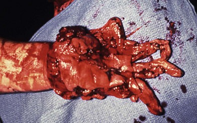
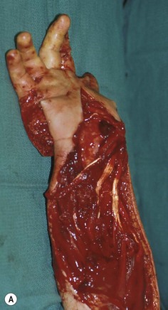
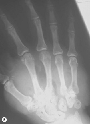
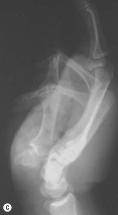
Avulsion injuries primarily involve the soft tissues of the upper extremity. The etiology of these injuries is often due to industrial accidents in roller press machines which both cause bone dislocations in the hand and can lead to loss of the entire soft-tissue envelope to the hand (Fig. 12.6). These injuries predictably lead to severe scarring and often poor function despite the type of reconstruction performed. Early motion is essential for reasonable outcomes in these injuries.


Roller injuries occur when the hand is caught between two rollers whose function is usually to compress sheets of metal. This can result in an array of injuries, including degloving of the skin of the hand, fractures, and potentially crushing of all the tissues of the hand and forearm. These are devastating injuries and call on the entire armamentarium of the surgeon to manage the bony fractures, dislocations, and coverage and functional issues from soft-tissue damage (Fig. 12.7).


Careful evaluation of both the patient and the injury, formulation of a treatment plan, meticulous operative treatment by an experienced team, and early, motivated rehabilitation reduce the morbidity associated with these injuries.


Diagnosis/patient presentation


Evaluation of the patient in the emergency room


Evaluation of the patient with a high-energy injury includes a thorough trauma workup, beginning with the basics of airway, breathing, and circulation. Whereas the mangled extremity is often the most apparent injury, careful evaluation of the entire patient for potential life-threatening or other associated injuries is critical to formulation of a treatment plan.


The patient’s history focuses on the time and mechanism of injury and any associated chemical, electrical, or thermal components of the injury. The mechanism of and the time from injury, which is especially crucial when ischemia is present, are the most important factors in determining the zone of injury and predicting the ability to salvage the extremity. A medical history is taken to determine the patient’s ability to tolerate a prolonged anesthetic with the potential for significant blood loss, fluid shifts, and release of metabolic byproducts after revascularization of ischemic tissue. Medical conditions such as diabetes, hypertension, vasculitis, or other inflammatory diseases, and smoking history can adversely affect outcome and should be considered in developing a treatment plan. Similarly, the occupational history and social history are important in determining postoperative compliance and in addressing the reconstructive goals. The presence of one or several adverse factors is not an absolute contraindication to salvage of an extremity or to microvascular repair and/or reconstruction. However, these factors should be considered in selecting the type of reconstruction to be used and in better predicting outcome.


Examination of the mangled extremity should be systematic and address vascular status, skeletal stability, motor and sensory function, and soft-tissue loss. The vascular status is evaluated by assessment of peripheral pulses, color, temperature, and capillary refill time in the distal extremity. Pulse oximetry is generally readily available in the emergency department and is helpful in assessing ischemia in the fingers. Doppler examination and angiography can also be used. Arteriographic exam in the mangled extremity may simply delay revascularization, however, and should be utilized very selectively. Skeletal injury is assessed clinically by the presence of deformity, crepitance, or bone tenderness. Radiographs should be taken of the entire extremity – particularly to evaluate the joints above and below the level of injury. A motor and sensory examination should be documented. The examiner should be aware that motor or sensory loss can result from muscle, tendon, or nerve injury as well as from ischemia. The ultimate assessment of the mangled extremity occurs in the operating room, however, after the debridement of nonviable tissue.


In the case of limb-threatening ischemia in a mangling injury, evaluation of the status of the vessels is generally performed in the operating room without the delay necessary for an arteriogram. An intraoperative angiogram may aid in determining the level and extent of arterial injuries, however. Radiographs taken in the operating room are usually of better quality than those taken in the emergency department because the extremity can be positioned without causing the patient discomfort. Traction radiographs allow better delineation of the fracture pattern and number of fragments, especially in evaluating intra-articular fractures about the wrist and elbow. Photographic documentation of the injury should be done throughout the course of treatment from initial evaluation to conclusion of treatment.9


Emergency room to operating room – making a plan


Planning in patient management starts when the patient is first seen in the emergency room. As a general rule, however, multiple examinations of the extremity by multiple physicians (emergency physician, intern, resident, fellow) are to be avoided. In the unanesthetized patient this is painful and leads to further anxiety which can lead to vasospasm and further problems. The quality of circulation in the extremity must be assessed, and a rough idea of the soft-tissue deficit should be obtained in a single examination. X-ray studies done in the emergency room of the extremity (and of the distal portion in cases of amputation) also can give an idea of what might be necessary in terms of bony fixation. Consent must be obtained for whatever might be necessary to include bony fixation, revascularization (including potential vascular graft donor sites), and coverage. While an emergency free flap is rarely indicated, the patient should be informed of potential soft-tissue flap donor sites as well. The patient and family should also be informed of the need for further surgery, including second looks/washouts which should be performed in the first several days.


Planning for reconstruction


In deciding how best to treat the mangled extremity, a variety of factors are considered. They can be broadly classified as patient and extremity factors. Pertinent patient factors include the general condition, age, handedness, occupation, functional requirements, and socioeconomic background of the patient. Associated injuries resulting in cardiopulmonary or hemodynamic compromise as well as pre-existing medical problems will mitigate against a lengthy salvage procedure, especially in a patient of advanced age. Conditions adversely affecting the blood vessels, such as diabetes mellitus, vasculitis, or smoking, will increase the risk of anastomotic failure and should be taken into consideration. Psychiatric disorders may be a contraindication to reconstruction because of possible repeated suicide attempts or anticipated noncompliance with the rehabilitation program. A morose patient may be temporarily incompetent to participate in determining treatment. Because time is a critical factor in treatment, it may be better to err in attempting to salvage an extremity than to perform a primary amputation.


Important extremity factors include the time since the injury, the severity of the injury, and the previous functional status of the extremity. Warm ischemia time longer than 6 hours results in progressively irreversible changes in cellular structure of muscle. Even if vascularity is re-established, tissue necrosis will not be avoided. Systemic risks of revascularizing a limb with prolonged ischemia must also be considered and addressed. These include acidosis, hyperkalemia, and rhabdomyolysis. In amputations of digits, where no muscle is present, the delay until reperfusion with use of cold ischemia may be extended to 20 hours. Finally, the previous condition of the extremity is considered. A history of major trauma, neurologic disease, or congenital deformity resulting in impaired function may not justify a salvage attempt.


The multitude of factors and the complex interrelations among them make reaching a decision a difficult task, even for experienced surgeons. Specialized scoring systems have been developed on the basis of lower-extremity injuries. These may offer valuable guidelines for evaluating the lower extremity but cannot be applied well to the upper extremity.10 Each case is unique, however, and the final decision should be an individualized one based on assessment of the patient and extremity parameters as well as sound judgment. The patient’s knowledge of the potential risks and benefits of surgery and the possibility of early or later amputation is important.


A third factor which is rarely discussed is the surgeon factor. The skill and experience of the surgeon managing the patient are extremely important in determining the outcome.11 While the experienced surgeon is more likely to have a more favorable outcome, reasonable results can be obtained by others if they follow basic principles and stick to what they are comfortable doing. The surgeon who only occasionally performs microsurgical tissue transfer may better serve the patient by performing a pedicled flap for initial coverage than attempting an esoteric and complex microsurgical procedure. It is always better to perform an initial reconstruction using a technique with which you are familiar rather than attempting to perform a procedure that has a high potential for failure. In any case, it is preferable to refer a patient with a complex extremity injury to a center which routinely deals with these problems rather than attempt reconstruction if one is not familiar with these types of problems.

Only gold members can continue reading. Log In or Register to continue

Stay updated, free articles. Join our Telegram channel

Feb 21, 2016 | Posted by in General Surgery | Comments Off on Reconstructive surgery of the mutilated hand

Full access? Get Clinical Tree

Get Clinical Tree app for offline access