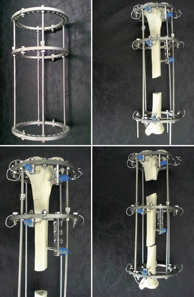
Fig. 9.1
The cutoff point values for the comparison of “amount of bone defect in cm” and “occurrence of complications” have been calculated using receiver operating characteristic (ROC) curve analysis (Istanbul Faculty of Medicine, Orthopaedics and Traumatology Archive)
9.2 Examination/Imaging
9.2.1 Physical Examination
The skin condition of the limb has an important role in the treatment. Viable skin envelope should be assured before bone reconstruction. When necessary, definitive treatment can be delayed or combined with soft tissue procedures.
Most of the patients with massive defects have vascular impairment due to previous injury or surgery. Vascular status assessment is vital, as the intervention itself may lead to additional injuries or insufficiency.
Proximal and distal joint range of motions and any existing deformity should be noted and corrected if necessary.
9.2.2 Imaging
True size anteroposterior (AP) and lateral radiographs are necessary for planning. In case there is deformity, a meticulous planning should be done before surgery with long-standing AP and lateral X-rays.
In case vascularized fibula graft is planned, both fibulae should be visualized by X-rays.
For osteomyelitis and tumor, magnetic resonance imaging (MRI) is mandatory. Extent of the resection and remaining bone stock has to be noted using MR, before surgery. MRI is also valuable for diagnosing skip metastasis (or skip abscesses) which will increase resection length and also for grading osteomyelitis.
If there is known or suspected vascular impairment due to previous surgery, current pathology, or trauma, conventional or computerized tomography, angiography, or Doppler ultrasound can be performed.
9.3 Surgical Anatomy
One must be familiar with all exposures possible in the limb, as the case may require any due to previous intervention. If it exists, the previous incision should be used to avoid any skin breakdown.
Any possible described exposure can be used. It should be individualized to the patient, by means of diagnosis, required neurovascular dissection, and preferred treatment method.
During surgery, the common peroneal nerve and both anterior and posterior tibial arteries are at risk.
Titanium cages require high amounts of graft, as it is used impacted. Both allografts and autografts can be used. In case autograft is preferred, posterior iliac approach should be preferred.
9.4 Positioning
Patient is prepared supine, with a sandbag under the ipsilateral buttock to prevent external rotation.
Prepare graft harvesting site if necessary.
Even if one fibula is planned for harvesting, both limbs should be prepared.
Fluoroscopy and radiolucent table are necessary throughout the operation.
9.5 External Fixation
9.5.1 Procedure (Segment Transport)
A circular external fixator is used for the segment transport technique. The frame consists of three rings applied to the same rods in order to allow sliding of the transported bone (Fig. 9.2).

Fig. 9.2
Schematic view of the segment transport method with a saw bone
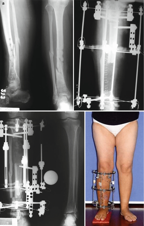
Technique: The resection area is determined by preoperative planning with X-rays. The frame should consist of three rings connected with the same rods as it allows sliding for segment transport. Following bone resection, the connective parts of the external fixator are assembled, required for either internal or external transport. If internal transport is planned, one of the rings is used as a “dummy” ring (no connection to the bone) for further compression of the docking site. However, we currently prefer doing the external transport technique due to its comfortability and easy facility. The osteotomy site for lengthening is prepared. First, the proximal reference wire is inserted and fixed to the most proximal ring. Then the distal reference wire is inserted and fixed to the most distal ring at the ankle level. Finally, all rings are fixed to the bone with Kirschner wires (K-wires) and Schanz pins (Fig. 9.3).


Fig. 9.3
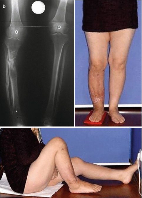
A fifty-one-year-old female patient with chronic osteomyelitis, who underwent a segment transport technique. (a) X Rays before surgery, during and after segment transport. (b) X Rays and knee flexion after fixator removal
Pearls
Pitfalls
Long waiting time for docking
Invagination of skin
Skin irritation and pain due to sliding bone
9.5.2 Procedure: Acute Shortening and Relengthening
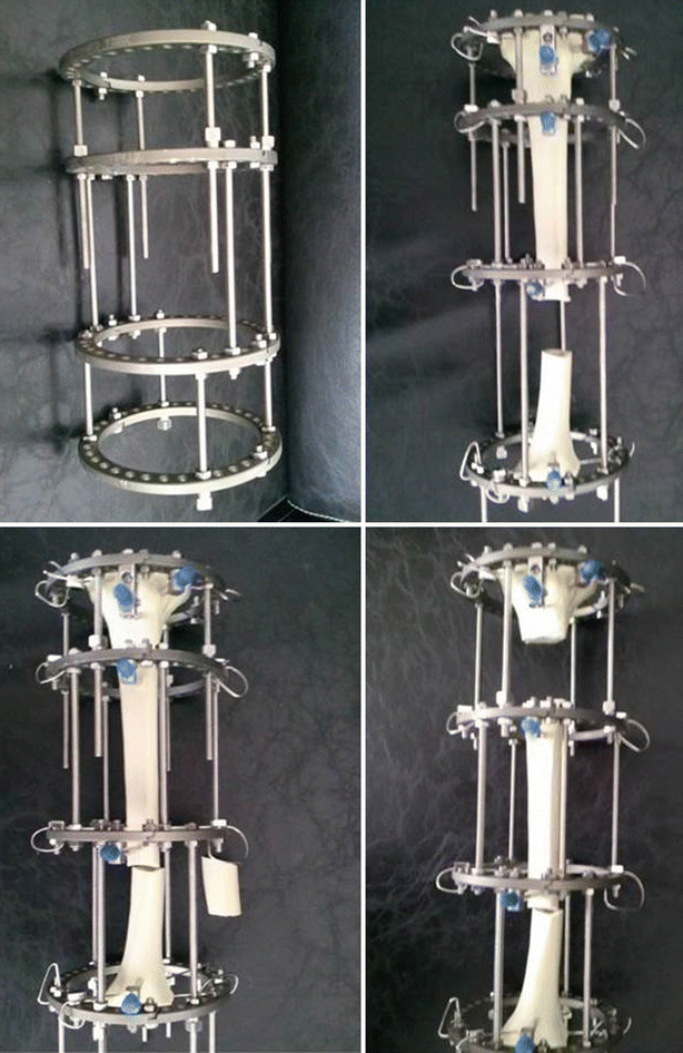
Preconstructed Frame: The circular external fixator is used as well for this technique. In this technique, the external fixator consisted of four rays; the rings were connected via different rods as needed. In the cases of distal tibial lengthening and short distal tibial fragments less than 4 cm, the frame is extended to include the foot in order to provide adequate stability (Fig. 9.4).

Fig. 9.4
Schematic view of the acute shortening method with a saw bone
Technique: The area of resection is determined by preoperative X-rays. A transverse incision is performed over the nonunion or fracture site and the resection is done. The resection of that site is done using an oscillating saw under saline irrigation to prevent heat necrosis. At this moment, the fibula is also resected not to prevent the docking of tibial bone ends. Moreover, the resection is performed until bleeding bone is achieved especially for nonunion cases. Then, acute shortening is done as long as the blood circulation of the foot exists. If the distal arterial circulation is maintained which is confirmed by capillary refilling, pulse oximeter, and Doppler ultrasound, respectively, as needed, the operation could be continued. If the circulation does not allow more shortening, gradual shortening should be reserved at 2 mm/day postoperatively. Following the shortening procedure, the skin is closed and the resected bone ends are temporarily fixed using K-wires or intramedullary Steinman pin. Then proximal and distal reference wires are inserted and fixed to the rings at the most proximal and distal level of the tibial bone. Then the middle two segments are fixed using two K-wires. At this stage, X-rays are taken on both planes. If the alignment of the tibia is well, the procedure goes on; otherwise, a malalignment on any plane should be corrected at this stage. Following this stage, fixation is completed at all levels of frame. Finally, an osteotomy is performed to relengthen the tibia at the healthy site of bone.


