Reconstruction of the soft tissues of the back

11 Reconstruction of the soft tissues of the back


Introduction


The back is 18% of the total body surface area, yet it is an area commonly neglected in older texts of plastic surgery. From the nape of the neck, to the anterior axillary lines, to the inferior gluteal crease, the only area well known to the plastic surgeon is the buttocks for the treatment of pressure sores. In years past, the common surgical procedures that involved the back were performed in areas of good vascularity and only uncommonly required procedures to aid in coverage. Small spine procedures, thoracotomy incisions, flank incisions for access to the retroperitoneum, and removal of soft-tissue tumors of the back such as lipomas did not inspire chapters in plastic surgery textbooks. What changed the equation were the advances made in spine instrumentation.


In decades past, spinal fusions were often performed by removing discs and scraping the end plates of vertebral bodies. These procedures were not limited by the surrounding soft tissues. This changed with improvements of surgical hardware that allowed for longer and longer constructs for spinal stability. Simultaneous anterior and posterior spine exposures for rigid fixation greatly stripped and elevated the surrounding soft tissues, and correspondingly pushed the soft tissues past the point where wound healing would be automatic. For illustration, the average wound in terms of vertebral body length in a 1995 paper was three bodies long, and many wounds were due to pressure sores of the back. By 2003, the average wound length was 11 vertebral bodies long, and all of the wounds had large amounts of hardware. In response to the need created by our surgical colleagues, surgical procedures to close the back reliably were developed.


Patient presentation


Midline back wounds


When a spine surgeon calls to discuss new drainage from a midline spine wound, the thought process for treatment should be methodical and thorough. When was the last procedure performed, and did the procedure involve the placement of hardware? What type of hardware is present, and is it locally prominent or is it low-profile? Did the spine surgeons place hardware on the anterior or lateral aspect of the vertebral bodies, or rather was it only posterior? Had there been any dural tears that were repaired, and do the spine surgeons have any evidence of a CSF leak? Do the plain films demonstrate hardware immediately deep to areas of wounds or drainage? Often a computed tomography (CT) scan is obtained, but hardware artifact is substantial and prevents an adequate evaluation of fluid collections.


After obtaining an understanding of what was done, an evaluation of wound-healing issues is necessary. Is the patient markedly malnourished and catabolic? Are obesity and dead space management problems? Had the patient been radiated for a spinal cord metastasis with the associated stiff and edematous soft tissues?


Next, an examination of the patient is often telling of what needs to be done. Continued soilage of dressings with fluid and dieback of the wound edges often point to deep fluid collections that are emerging from around the spinal hardware. Is the area of wound breakdown at a pressure point due to incomplete restoration of spine architecture? Further understanding of the time course of the drainage also points to the underlying pathology. Persistent postoperative fevers ascribed to “atelectasis” are a common diagnosis made for these patients.


The time course of the presentation is critical. Early postoperative episodes of drainage less than 4–6 weeks after the spine surgery are typically successfully treated with repeat surgery and soft-tissue reconstruction with hardware preservation. However, drainage that has been only partially treated with a small debridement or treated solely with intravenous antibiotics, only to resurface months later, is more difficult to treat. In fact, a chronic hardware infection defined as bacteria in association with hardware greater than 6 months after placement is typically not a situation that plastic surgery can definitively treat without hardware removal.


Much can be learned with the simple examination of a patient’s chest X-ray in terms of the existence and location of hardware. Otherwise, plain films of the spine are obtained to reveal the length of the construct when present, degenerative spine disease, and the presence or absence of fusion. CT scans and magnetic resonance imaging (MRI) are helpful to look for fluid collections, pseudomeningoceles, and inflammation of the soft tissues. A key issue is if fluid collections are above or below the standard back muscle closure. If fluid or hematoma is seen deep to the musculature, a deep hardware infection is more likely. Unfortunately, the spinal hardware causes much artifact of both CT scans and MRI, lowering their ability to demonstrate fluid collections with high accuracy.


Patient treatment


Local wound care


For superficial, relatively painless wounds without exposed hardware, local wound care with dressings is a relatively risk-free way to achieve wound closure. Draining midline incisions tend to have wide areas of undermining along the length of the closure. These tunnels can typically be opened in the office with local injection of anesthesia and finger fracturing of the incision. A long wound without tunnels with a saucer shape typically heals faster than small wounds with a fishbowl-shaped internal wound due to the improved ability to cleanse the surface of the internal wound. Therefore, tunnels should be opened along old incision lines. All necrotic tissue should be debrided. All nonabsorbable sutures such as braided polyester should be removed. Obtaining local wound control with wide exposure of the wound is a tried and true method as the first step in healing. Patients are often unaware of the real size of the wound, and must be prepared for the resulting appearance of the surgical site after opening of tunnels.


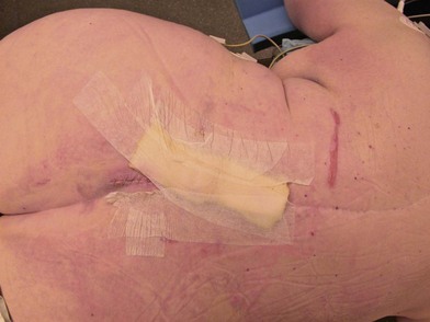
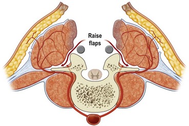
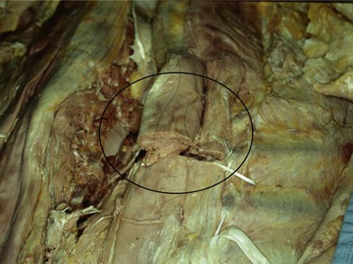
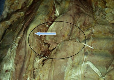
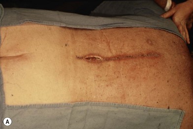
Much depends on the patient’s home situation. Dressings can be simple wet-to-wet saline dressings, with twice-a-day showers to cleanse the surface of the wound. Subatmospheric-pressure dressings can be employed, but the tubing is sometimes difficult to place under back-bracing devices (Figs 11.111.3). After a wound has granulated, if it is painful, then delayed primary closure or skin grafts can be performed if the wound is sizeable. However, the risks of these secondary closure procedures often outweigh the benefits. The subcutaneous tissue stiffens during the time of obtaining local wound control, and it is difficult to re-elevate and close without a fairly sizeable procedure.


Operative debridement


Patients with persistently draining wounds after spine surgery associated with hardware should be evaluated critically for an operating room debridement. Secondary indications for a debridement include unexplained fevers and fluid collections seen on imaging scans. The patient should be readied for surgery with large intravenous lines and in a warm operating room. The large exposed surface areas can allow the patient to cool rapidly, with associated coagulopathies and increased blood loss. Blood should be available for a transfusion. Due to the segmental nature of the vascularity, surgery does not typically expose large and few blood vessels, but rather many small ones, and the blood loss is rather constant throughout the case.


The maneuvers in the operating room are a critical step in the treatment of patients with postsurgical back wounds. A thorough incision and drainage should be performed for patients with unexplained or purulent drainage through their incision. The entire length of the incision should be opened as widely as necessary to explore for purulence and drain fluid collections. The erector spinae muscle closure should be reopened for cultures and to evaluate for liquefying hematomas. The surgeon should be knowledgeable at the beginning of the debridement if a laminectomy had been performed at the original spine surgery in order to prevent injury to the spinal cord and dura during the incision and drainage.


At this point, the surgeon will need to make a decision as to the quality of the tissues. Nonpurulent benign fluid collections in the subcutaneous tissues with no purulence deep to the musculature can be reclosed over drains, or else closed secondarily with a subatmospheric-pressure dressing (see above). Purulent and deep collections require additional decisions. If the local wound is so purulent as to preclude an immediate reclosure, then all nonviable tissue should be debrided, the wound irrigated, and left open for local wound care. This can be done with dressing changes on the floor for several days until a second trip to the operating room. Alternatively, a subatmospheric-pressure dressing may be applied, but this often involves a return trip to the operating room for its next exchange.


For those deep wounds judged amenable to reclosure, a radical incision and drainage are performed. This should involve the surgical excision of scarred tissues where possible to reveal supple soft tissues with pulsatile bleeding. Scar, though it bleeds, does not bleed in pulsatile fashion due to the small size of the vessels that developed during prior wounding. Pulsatile bleeding at wound margins has been shown to correlate with wound healing in problematic incisions, such as distal foot amputations. Tissues with a pseudobursa should be excised, as this too represents scar. Tissue that is stiff is unyielding and does not conform well, and so the tissues should be removed until they are soft to palpation. Pulsatile irrigation with saline is gentle to tissues that cannot be removed and yet still is able to cleanse surface bacteria. A stronger debriding instrument has been developed (Versajet, Smith and Nephew) that can remove a thin layer of tissue at a time with pressurized streams of sterile fluid and this is effective in these large open wounds.


An interesting issue is the removal of nonviable elements such as hardware and bone graft. While plastic surgery teaching emphasizes the removal of all nonvascularized surfaces, in this case the hardware acts to stabilize the wound. An orthopedic principle is that the most effective way to fight infections is to have rigid bone fixation. Therefore, hardware that is well fixed should remain in place in early hardware infections. This is done both to stabilize the wound for improved healing as well as to avoid the surgery involved with removal and later replacement of hardware. Long-term maintenance of the hardware as well as clinical and radiographic evidence of fusion are well documented in patients who were returned to the operating room for washouts within 6–8 weeks of placement. As stated previously, hardware colonized after 6 months is defined as a chronically infected foreign body, and salvage of this hardware is much less likely long-term. Another issue is the endogenous and exogenous bone graft placed to achieve a fusion. In the absence of definitive studies, it seems reasonable to remove nonincorporated and easy-to-remove graft, but to leave in place graft that has in any way begun to stick to the local tissues due to inosculation.


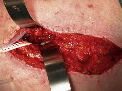
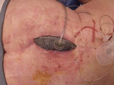
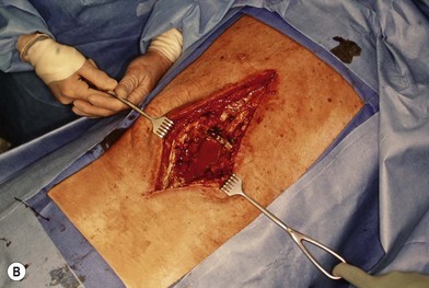
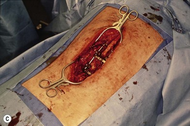
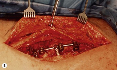
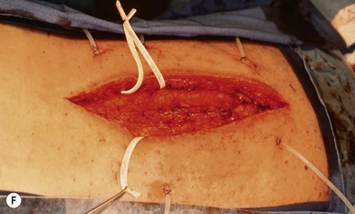
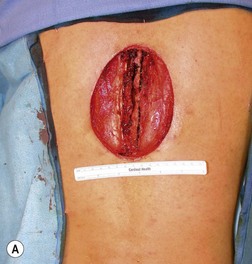
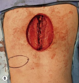
Wound “shape” is an important concept for reconstruction of the soft tissues of the back. The back has lordotic and kyphotic regions in the normal condition, and these contours can be dramatically changed with pathology. This serves to make postoperative positioning difficult when the surgical incision is also the most prominent portion of the back. The depth of the procedure performed by the spine surgeons is also important. A patient with a laminectomy by definition has a deeper wound than when the spinous processes are intact. Spinal hardware serves to create a local prominence in contradistinction to the depth of the surgical dissection. A prime reason for persistent fluid collections is the three-dimensional space between a laminectomy and the adjacent vertically oriented bars. Crosslinks between the bars further prevent soft tissue from collapsing into the space immediately over the midline. Finally, laterally placed hardware can be poorly covered by soft tissue and may be the originators of pressure sores. This is especially common when hardware inserts into the posterior superior iliac spines for pelvic fusions. Therefore, in the treatment of wounds of the spine, the three-dimensional shape should be evaluated and converted as much as possible to a two-dimensional wound. Prominent hardware should be exchanged for something with a lower profile (Figs 11.411.7). Patients with incomplete corrections of the spine deformities should be revised to recreate better the natural contours. The deeper the hole, the more a flap should be “dropped into” the defect, rather than tissue simply slid towards the midline. This requires a more redundant soft-tissue flap such as an omentum to fill the defect appropriately. Teamwork between the spine and soft-tissue surgeons is often necessary to treat problems of wound shape and contour appropriately.
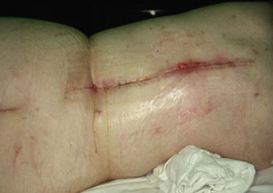
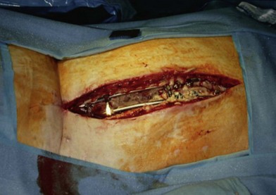
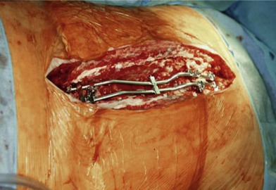
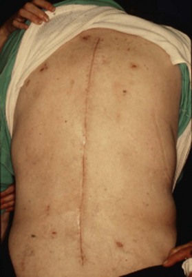
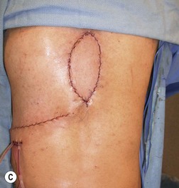


The status of the CSF and the dura is also critical for treatment of certain back wounds. The dura is opened and closed in a planned fashion in numerous instances, including the treatment of spinal cord malignancies, untethering of the spine for patients with a history of cord lipomas and spina bifida, and treatment of pseudomeningoceles. Unplanned tears of the dura also occur in laminectomy procedures. The low-pressure CSF leaks and the continuing fluid buildup push the soft tissue away from the leakage site, and thereby prevent soft-tissue collapse of the space. The CSF fluid in fresh postoperative cases can also become infected, and exist in association with surgical wounds of the back. Treatment of patients with CSF leaks will be discussed later in this chapter.


Flap closure


Principles


The first step in reconstruction is a timely debridement, as has been emphasized already. The second step is local wound control with a radical debridement of all stiff and scarred tissue. For the reconstruction, the surgeon should never rely on scar to be a positive factor for wound healing. The more completely the old scar can be excised and replaced with nonscarred new soft tissues, the better the reconstruction. Finally, the reconstruction should be performed to do the “maximum for the minimum.” The procedure with the highest chance for success and with the lowest morbidity should be selected for the patient.


Central questions to be answered include the presence or absence of a fusion, and the vertebral levels involved.1 When an instrumented fusion has been performed, then the erector spinae musculature function is no longer necessary, and the muscles are completely expendable in terms of a reconstruction. The fusion rods prevent postoperative motion, and so when the erector spinae muscles are reapproximated in the midline, they tend to stay there. Spine patients without fusions still require the function of the erector spinae musculature when healing is completed for flexion and extension of the spine. These patients do better with flaps that are “dropped into the hole,” rather than with erector spinae muscles that are closed side to side and that would dehisce with back flexion.


Local wound care often suffices for superficial wound problems above the erector spinae. After the deep aspect of the wound is judged to be without purulence or fluid collections, a subatmospheric dressing can be applied, often with a benign postoperative course. This does well for obese patients with thick subcutaneous tissues. Delayed closures are possible after the wound has granulated, but the failure rate in heavy patients where normal body movements pull against the suture line is substantial. Subatmospheric-pressure dressings are reported to allow soft tissues to cover over small areas of exposed hardware, but this has not been my experience.


Possible flap choices for spine closure


Erector spinae muscle flaps


The erector spinae muscles, also called the paraspinous muscles,2 are expendable after a previous spine fusion and no longer are functional for spine extension and flexion. The dissection of the muscles proceeds in stepwise fashion in order to move the muscles towards this midline. This flap is appropriate from the high cervical area to the low lumbar area, but it will not adequately cover an occipitospinal fusion, and nor will it be sufficient for lumbosacral soft-tissue coverage. One must be careful in its use when a lateral approach to the spine has been made, because the muscle can be transected for access.


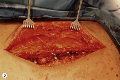
First, skin flaps are elevated superficial to the thoracolumbar fascia (Fig. 11.8). With a retractor putting tension on the soft tissues, with cautery the granulation tissue is entered, and with finger dissection a blunt plane is elevated. The latissimus muscle and trapezius muscle should stay attached to the skin. The dissection is easiest inferior in the lumbar area where the muscle is round and large, and most confusing superiorly where the muscle is thinnest and becomes attached to the undersurface of the trapezius. The erector spinae muscles have a convex shape, and there is a rounded aspect of the muscle that then descends laterally towards the more lateral neck, thoracic, and lumbar areas. It is in this groove that segmental blood vessels enter the lateral and deep aspects of the longissimus and iliocostalis muscles. Continuation of these blood vessels continues more superficially to the skin and to the latissimus muscle in the thoracolumbar area. While the surgeon is elevating the skin flaps, the perforators and dorsal sensory nerves going up to the skin often can be identified and preserved to maintain skin vascularity. To lessen the chance of marginal skin perfusion and to limit a future potential seroma cavity, undermining should not be overdone.


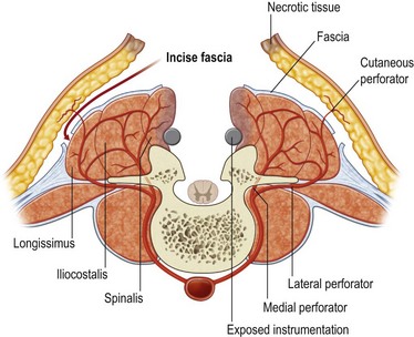
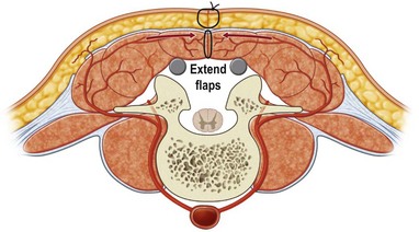
The thoracolumbar fascia is incised much like the development of any standard bipedicled flap in order to mobilize the erector spinae muscle and/or overlying skin to the midline. When the original dissection is superficial to the fascia and the fascia remains attached to the muscle, this facilitates the later closure, and the fascial release is critical to the movement of the muscle. Occasionally the fascia remains attached to the skin during the initial elevation. In this case, muscle mobilization is facile, but the skin is more difficult to close in the midline. Therefore, an incision of the fascia along its length allows the skin to move medially and to provide the excess for debridement and reclosure. The dissection up until this point is rather easy and bloodless, but only moves the muscle an estimated 30% of its potential. Complete mobilization of the muscle requires a dissection along the deep aspect of the erector spinae (Fig. 11.9). Again with tension applied on to the tissue with retractors, cautery dissection of the deep and medial attachments of the erector spinae will mobilize the muscle and detach it from the lateral aspects of the transverse processes of the spine. It will by necessity divide the medial row of blood vessels entering the paraspinous muscles, but the prior dissection to identify the lateral vessels entering the muscle will suffice to preserve vascularity. One technique is to have the fingers of the nondominant hand in the lateral groove at the site of the lateral perforators while bluntly elevating the medial aspect of the muscle with the thumb and index finger. The medial muscle elevation is a powerful means to allow the paraspinous muscles to “unfold” like an accordion to be advanced toward the midline (Figs 11.10 and 11.11). The muscle changes shape from being a round muscle mass to being more elliptical. Advancement of the muscle is a process much different from simply releasing the thoracolumbar fascia and “rolling” the dorsal aspect of the muscle toward the midline.


With the skin and muscle flaps so created, it is now quite easy to distinguish stiff and inflamed tissue from more normal soft tissue. The medial aspect of the skin, subcutaneous tissue, and paraspinous muscles is sharply debrided. The muscles are brought together in the midline (Fig. 11.12), and any extra tissue can be imbricated to help fold the soft tissue into crevices between vertically oriented hardware bars (Fig. 11.13). Drains are left both deep and superficial to the erector spinae closure, and are left in until the drainage is minimal. When the erector spinae muscle closure is of good quality, there is no need for a second overlying muscle flap using the trapezius or the latissimus muscles.


Latissimus muscle or myocutaneous flap


The latissimus muscle is a well-known and understood flap.3 The donor site is minimal. Based on the thoracodorsal pedicle, the muscle can be moved superiorly up to the level of the top of the scapula. Based on its minor perforators that also supply the paraspinous muscles, the latissimus can reach the lower lumbar area. An advantage to the latissimus is being able to be “dropped in” to a hole, and so it is useful for patients who have not been fused, or for more lateral defects. The muscle can reliably carry a skin paddle, and this often helps in the inset of the flap. Caution should be taken in patients who have had thoracotomy incisions, as the muscle is often divided.


The latissimus muscle is a good but still second-line flap for the coverage of midline back wounds. In comparison to the paraspinous flaps, the latissimus flap can only cover a spine wound 10–12 cm in length, while the erector spinae flaps can cover practically the entire length of the spine. The dissection required to elevate the latissimus muscle takes more time and effort than does the erector spinae. While low, there is a donor defect from the loss of the latissimus muscle, especially in patients who may need assistive devices to ambulate. The latissimus flap is a good choice for nonfused wounds of the back (Fig. 11.14), or when the erector spinae muscles have been radiated.


Stay updated, free articles. Join our Telegram channel

Feb 21, 2016 | Posted by in General Surgery | Comments Off on Reconstruction of the soft tissues of the back

Full access? Get Clinical Tree

Get Clinical Tree app for offline access