Assessment of injury
Successful long-term reconstruction of the burned lower extremity requires attention paid to both form and function. The limb is responsible for upright positioning, weight bearing, and ambulation, and it is a critical part of the larger musculoskeletal system. As a result, lower-extremity disability has a significant impact on quality of life. Cosmesis of the leg is also important given its frequent exposure in clothing, and revision surgery for aesthetic concerns is common in limb salvage. , The burned lower extremity can be evaluated systematically by subdividing it into its tissue types.
Skeletal assessment
Concomitant thermal and bony injury is common. Timely skeletal evaluation with physical examination and imaging is important. Treating fractures early after burn is both safe and effective. , It ensures skeletal stability, permitting safe wound care and early physical therapy. Orthopedic open reduction and internal fixation may be performed if the surgical approach is through unburned tissue. External fixation is recommended if there is concern for soft-tissue viability to limit risks of infection, exposure, or nonunion. For severe injuries that permanently compromise weight bearing or joint range of motion, salvage may be unreasonable. A team-based approach is recommended and results in superior outcomes.
Neurovascular assessment
Neurovascular integrity in the lower extremity is important for short- and long-term limb salvage. Initial evaluation of an extremity burn should always include assessment of neurologic function and careful documentation of peripheral pulses. Circumferential injuries require regular reevaluation to permit early identification of dysfunction.
Compartment syndrome results from elevated pressures within a rigid anatomic space. This can be due to surrounding inelastic burned soft tissue, direct tissue damage from electrical burn, or mixed mechanism of injury. As these pressures exceed diastolic pressure, perfusion is compromised resulting in limb ischemia. Compartment syndrome is characterized by five classic signs and symptoms—pain, paresthesia, pallor, paralysis, and pulselessness—and prompt diagnosis requires clinical acumen or direct measurement of compartment pressures with the appropriate device. A worsening neurovascular exam, like diminished amplitude of palpable pulses, may also indicate increasing compartment pressures. Early escharotomy and/or fasciotomy with preservation of vulnerable superficial anatomy is required to preserve or restore perfusion to the distal extremity ( Figs. 67.1–67.3 ). Failure to identify loss of extremity perfusion can result in acute limb loss or chronic limb dysfunction.
Thigh fasciotomy.
(Artist credit Erin Wolfe, MD.)
Lower-leg fasciotomy.
(Artist credit Erin Wolfe, MD.)
Lower-extremity fasciotomies with relevant superficial anatomy.
(Artist credit Erin Wolfe, MD.)
Peripheral pulses may also be diminished due to underlying premorbid vascular disease. For patients with history or exam consistent with this diagnosis, evaluation of the extremity with objective vascular studies is recommended. Toe pressures serve as an excellent noninvasive indicator of peripheral vascular status and are largely unaffected by vessel calcification. Low toe pressures suggest poor wound healing. Arterial profile uses ultrasonography to identify specific areas of stenosis. Abnormal study results should lead to a vascular surgery consult, and flow should be optimized before attempts for surgical wound closure.
Peripheral nerve dysfunction occurs in both acute and chronic phases of burn care. Neuropathic pain is the most common early sign of nerve injury. Symptom control with medication is the mainstay of treatment, although fat grafting appears promising. Peripheral nerve compression is a late finding and may be treated with targeted surgical decompression. Although an insensate foot has long been considered an indication for amputation, recent studies have called this into question.
Soft tissue assessment and maintenance
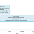
Physical examination of the wound serves as the primary method of assessing burn depth and healing potential. Superficial (first-degree) burns are isolated epidermal injuries that are dry, red, and painful. Partial-thickness (second-degree) burns involve epidermis and partial dermis, blister, and are subdivided by depth. Superficial partial-thickness burns will blanch, whereas deep partial-thickness burns will not. Indeterminate middermal burns can prove challenging to predict and treat, so frequent reevaluation is recommended. Full-thickness burns injure both epidermis and dermis and can extend into deeper tissues, and are characterized by dry, leathery, insensate eschar. Resuscitation, infection prevention, nutrition, and local wound care are critical to optimize healing potential of partial-thickness burns. When possible, a transition to nondaily dressings improves pain control and promotes mobility. Physical therapy is initiated early, often within 24 hours, to ensure that joints remain supple with full range of motion. Ultimately, surgical debridement serves as the best way to define the true soft tissue defect.
Acute phase reconstruction
Acute lower-extremity reconstruction focuses on closing wounds while optimizing functional status during the healing process with the goal of a normally functioning extremity. Superficial partial-thickness burns can be allowed to heal by themselves with local wound care alone. Middermal and mixed partial-thickness injuries may heal without surgery or may require excision and coverage, usually with an autograft. Spray-on autologous skin cell suspensions are an emerging way to facilitate healing in these cases while minimizing donor-site morbidity and allowing for ongoing active physical therapy. Deep partial-thickness burns that do not exhibit healing potential and full-thickness burns are managed with tangential excision and, ultimately, autografting. Most smaller injuries can be managed in a single stage. Compared with sheet grafts, mesh grafts offer improved take and reduce donor-site size but produce more contraction and worse cosmesis. , Unexpanded mesh grafts offer a reasonable compromise, especially over joint surfaces. When the injury is large or the wound bed perfusion is difficult to assess, a staged approach with temporary coverage with a skin substitute may be indicated before autografting.
Loss of autograft on the dependent surfaces of the proximal posterior thigh and buttock is common. This is due to a combination of factors, including shear during position changes, localized ischemia from pressure, and proximity to the anus and genitals. Limited areas can be allowed to heal secondarily, whereas large areas of graft loss may require regrafting. Regardless, in patients with anterior and posterior burns, prioritizing the use of the best segments of autograft for anterior injuries is recommended.
In deep burn injuries, ungraftable tissues (e.g., tendon or bone) may be encountered after debridement. For limited areas or in patients unable to tolerate long surgeries, bioengineered skin substitutes with staged skin grafting are useful. Thermally damaged, necrotic tendons at the ankle may simply be resected, permitting wound contracture with aggressive physical therapy to fuse the ankle in a functional position.
Open joints, exposed neurovascular structures, or large areas of exposed viable tendon or bone, however, may require flap reconstruction ( Table 67.1 ). Exposed femoral vessels are covered with local flaps like the sartorius, the gracilis, and the vertical rectus abdominis myocutaneous flap. Abundant soft tissue in the thigh means flap coverage is not commonly required. The approach to lower leg reconstruction divides the region into thirds. The proximal third, including the knee, is reconstructed with gastrocnemius muscle flaps, although genicular artery perforator flaps are viable options for smaller defects and obviate the need for skin grafting. Middle-third defects are addressed with a soleus muscle flap. The distal third has limited local options. Reverse soleus or sural flaps, perforator flaps based on anterior or posterior tibial vessels, or bipedicled flaps all may be used, although care must be taken to design flaps based on vessels uninjured by the burn. Local flap reconstruction of the distal third can be challenging and unreliable. Free tissue transfer is a powerful reconstructive tool, even in the acute phase.
Table 67.1
Reconstructive Options for the Lower Extremity by Anatomic Region
| Anatomic Region | Reconstructive Options |
|---|---|
| Hip |
|
| Knee |
|
| Ankle |
|
| Foot |
|
Related posts:


Stay updated, free articles. Join our Telegram channel

Full access? Get Clinical Tree


