Fig. 61.1
Localized pagetoid reticulosis. A solitary, slowly enlarging, persistent erythematous patch with sharply demarcated borders for 10 years
Pathology
Histologically, LPR demonstrates a hyperplastic epidermis with striking colonization by atypical lymphocytes, especially in the lower reaches (Fig. 61.2). The lymphocytes are distributed singly, in small clusters or in large lacunae (Fig. 61.3). Involvement of adnexal epithelium is often a feature. The lymphocytes are medium to large, with hyperchromatic irregular nuclei and abundant vacuolated cytoplasm (Fig. 61.4). They usually have a perinuclear halo. Mitotic figures are sometimes conspicuous. The papillary and superficial dermis shows a mixed cell infiltrate with very sparse atypical cells.
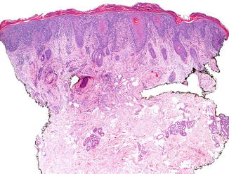
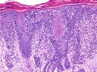
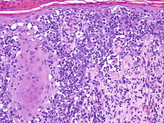
Fig. 61.2
Localized pagetoid reticulosis. This biopsy of an acral lesion shows an acanthotic psoriasiform epidermis covered by hyper-orto and parakeratosis and permeated by numerous lymphocytes
Fig. 61.3
Localized pagetoid reticulosis. The lymphocytes are disposed singly, in small clusters and in large lacunae at all levels of the epidermis, but especially in its lower reaches
Fig. 61.4


Localized pagetoid reticulosis. The lymphocytes are medium to large with hyperchromatic, irregular, haloed nuclei
Related posts:


Stay updated, free articles. Join our Telegram channel
Full access? Get Clinical Tree
