Psoriasis
The clinical presentations of psoriasis listed here create a somewhat artificial classification, which is based on the characteristics or morphology of the predominant type of lesion and its distribution. There may be composites of these different types in a given patient. Because each type is managed somewhat differently and has its own differential diagnosis, each is discussed separately.
 Localized plaque psoriasis
 Generalized plaque psoriasis
 Acute guttate psoriasis (see also Chapter 27, “Special Considerations in the Skin of Pediatric and Elderly Patients”)
 Erythrodermic psoriasis (exfoliative dermatitis secondary to psoriasis) (see also Chapter 25, “Cutaneous Manifestations of Systemic Disease”)
 Psoriasis in children (see also Chapter 27, “Special Considerations in the Skin of Pediatric and Elderly Patients”)
 HIV-induced psoriasis (see also Chapter 24, “Cutaneous Manifestations of HIV Infection”)
 Inverse psoriasis (on the groin, penis, axillae, and perianal and inframammary regions)
 Psoriasis of the palms and soles
 Scalp psoriasis
 Psoriatic nails (see also Chapter 13, “Diseases and Abnormalities of Nails”)
 Psoriatic arthritis
 Other rare clinical variants: localized pustular psoriasis (Hallopeau) and the rare generalized pustular psoriasis of von Zumbusch
Overview
Psoriasis (psoriasis vulgaris) is a red and scaly chronic skin condition of unknown cause. The primary concern to most patients is the unsightly appearance of lesions whose visibility and persistence often lead them to feel self-conscious and unclean. The emotional toll and the personal struggle to come to terms with psoriasis are expressed in an autobiographic short story, “At War with my Skin,” by John Updike, an author with severe psoriasis. After undergoing an operation for a broken leg, Updike reflected, “I chiefly remember amid my pain and helplessness being pleased that my shins, at that time, were clear and I would not offend the surgeon.”
Psoriasis affects 1% to 2% of the world’s population. The condition is much less common in West Africans, African-Americans, Native Americans, and Asiatic people than it is in whites. It is found equally in men and women. Psoriasis most frequently begins in the second or third decade of life, but it can first present in infants or in the elderly. About 30% of patients with psoriasis have a family history of the disease. Patients may also develop psoriatic arthritis, which may precede or follow the onset of skin lesions.
Psoriasis can be a major blow to one’s ego. It may stifle social activities and sexual spontaneity, interfere with job opportunities, and inhibit participation in sports and the use of beaches and public swimming pools. Because psoriasis is a visible disease, it may arouse a fear of contagion, as well as repugnance and avoidance, from persons who are not used to seeing it. The young child with psoriasis has the additional burden of embarrassment caused by the undisguised scrutiny and thoughtless remarks of other children. A parent may have to cope with guilt for having genetically passed psoriasis on to his or her child.
A person who has psoriasis often spends an excessive amount of time treating skin lesions and trying to hide them, as well as searching for external causes and possible cures. The National Psoriasis Foundation provides information about psoriasis to educate patients, the public, and health care providers. The contact information is as follows: 6600 S.W. 92nd, Suite 300, Portland, OR 97223; 800-723-9166; www.psoriasis.org.
Pathophysiology
Psoriasis is essentially an inflammatory skin condition with abnormal epidermal differentiation and hyperproliferation. It is suggested that the inflammatory process is immunologically based and most likely set off—and maintained by—T cells in the dermis.
The lesions of psoriasis result from an increase in epidermal cell turnover. The cell’s transit time from the basal layer of the epidermis to the stratum corneum is decreased from the normal 28 days to 3 or 4 days.
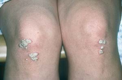
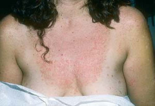
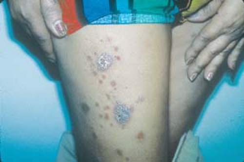
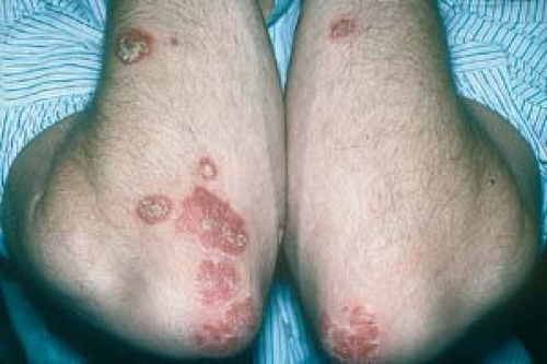
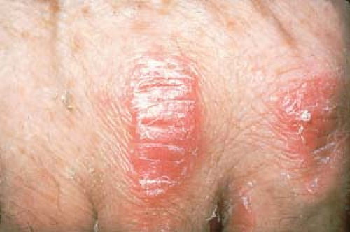
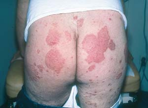
This “turned-on” epidermis, with its rapid accumulation of cells, accounts for the characteristic lesion of psoriasis: a red papule or plaque (Fig. 3.1). It also explains the accumulation of white or silvery (micaceous) scale; the great increase in cellular kinetics does not allow time for shedding (Figs. 3.2 and 3.3).
Because psoriasis is now considered to be an immunologic disease, most current therapies, including topical corticosteroids, phototherapy, photochemotherapy, methotrexate, and cyclosporine, are directed at the suppression of responsible T cells.
 3.1 Psoriasis. This is a typical location for the characteristic lesions of psoriasis. Note the well-circumscribed erythematous plaques surmounted by a fine scale. |
Histopathology
The histopathologic findings demonstrate the altered cell kinetics of psoriasis (Ill. 3.1):
Increased mitosis of keratinocytes, fibroblasts, and endothelial cells. Skin biopsies typically show:
Marked thickening (acanthosis) and also thinning of the epidermis with resultant elongation of the rete ridges
Parakeratosis (nuclei retained in the stratum corneum)
Inflammatory process:
Dermal inflammation (lymphocytes and monocytes)
Epidermal inflammation (polymorphonuclear cells) in the stratum corneum that may form the so-called microabscesses of Munro
Distribution of Lesions
The distribution of thickened, reddened, silvery or whitish, scaly papules or plaques can range from only a few small asymptomatic lesions on the elbows and knees to larger plaques that cover extensive areas of the body.
Psoriasis tends to be remarkably symmetric. It usually spares the face. Lesions are most commonly located as follows:
On large extensor joints (elbows, knees, and knuckles)
On the scalp
On the anogenital region (perineal and perianal areas, glans penis)
On the palms and soles
Trunk lesions may be small, guttate (teardrop-shaped) plaques or large plaques.
When psoriasis involves only the scalp and retroauricular areas, it is sometimes referred to as “sebopsoriasis” or “seborrhiasis” (see Figs. 2.56 and 2.58).
When the entire body is involved, generalized, disseminated plaques or exfoliative erythroderma may be evident.
When lesions occur primarily in the intertriginous areas (inguinal creases, axillae, and inframammary, perineal, and perianal areas), this manifestation is referred to as inverse psoriasis.
Psoriasis is commonly a cause of nail deformity, which is often mistaken for, and treated incorrectly as, a nail fungus infection (onychomycosis).
Psoriatic arthritis occurs in 5% to 10% of patients who are diagnosed with psoriasis.
Clinical Manifestations
Pruritus
Psoriasis generally is asymptomatic, but it can become quite pruritic and uncomfortable, particularly during acute flare-ups or when it involves the scalp or intertriginous regions.
The Köbner Reaction (Isomorphic Response)
Patients commonly recognize the phenomenon that new lesions may appear at sites of injury or trauma to the skin.
Fissuring of Plaques
Painful fissures may occur when lesions are present over joints, intragluteally, or on the palms and soles.
Psychosocial Problems
The health care provider should be attuned to the psychologic ramifications of psoriasis—anxiety, social isolation, alcoholism, depression, suicidal ideation—as possible associations and outcomes of this essentially benign skin disease. Stress has been implicated in the acute exacerbations and progression of psoriasis. In a vicious cycle, the poor self-image that may be incited by lesions can create more stress. Factors that may adversely influence psoriasis include:
Alcohol. Alcohol overindulgence has been reputed to exacerbate psoriasis, which, once again, can create a vicious cycle of worsening the alcohol overindulgence.
Drugs. Antimalarials, beta-blockers, angiotensin-converting enzyme inhibitors, certain nonsteroidal anti-inflammatory drugs (e.g., indomethacin), systemic interferon, and lithium carbonate have been reported to worsen psoriasis; however, preexisting psoriasis is not necessarily a contraindication to their use. Both systemic and potent topical steroids have been known, albeit rarely, to trigger a severe, acute, potentially fatal pustular psoriasis (pustular psoriasis of von Zumbusch) that tends to occur after withdrawal of the medication.
Physical trauma. Surgery, thermal and chemical burns, and infections potentially exacerbate psoriasis. For example, an increase in psoriasis activity has been observed in patients who are, or become, infected with the human immunodeficiency virus (HIV).
Sunlight. A small minority of patients find that their psoriasis is worsened by strong sunlight, probably via the Köbner reaction (Fig. 3.4), whereas the vast majority of patients generally consider sunlight to be beneficial.
Other factors. There is evidence that psoriasis is associated with smoking, obesity, and dyslipidemia. It has also been noted that severe psoriasis appears to increase the risk of a myocardial infarction.
Course
Psoriasis is an erratic condition with an unpredictable, waxing-and-waning course. It has no known cure; however, there are many methods of keeping it under control.
Many patients tend to improve during the summer and worsen during the colder periods of the year. This fluctuation is presumably the result of the positive influence of sunlight on psoriasis.
There have been anecdotal reports of patients with psoriasis who were “cured,” and some patients claim that they “grew out of it.” This situation may be explained by either a misdiagnosis of the original skin problem or by cases of acute guttate psoriasis (see later discussion) that resolved without recurrence.
The differential diagnosis of psoriasis varies, depending on the type and location of lesions. In general, psoriasis most often must be differentiated from the following:
Eczematous dermatitis
Superficial fungal infection
Seborrheic dermatitis
Pityriasis rosea
Drug eruption
Parapsoriasis and mycosis fungoides (cutaneous T-cell lymphoma)
Secondary syphilis, the “great imitator”
Diagnosis
The diagnosis of psoriasis is made on clinical grounds.
A skin biopsy or fungal studies may be performed to rule in or rule out other possible diagnoses.
Localized Plaque Psoriasis
Basics
In its mildest manifestation, psoriasis is an incidental finding and consists of mildly erythematous, scaly patches on the elbows or knees. Localized plaque psoriasis, the most common presentation of psoriasis, may remain limited and localized (Figs. 3.6, 3.7, And 3.8), or it may become unstable and become widespread.
Diagnosis
If the typical well-demarcated whitish or silvery plaque is present in the usual locations, the diagnosis of psoriasis is quite evident.
Other helpful diagnostic features include a family history of psoriasis and nail findings (see later in this chapter and also Chapter 13, “Diseases and Abnormalities of Nails”).
If necessary, other tests, such as a skin biopsy and fungal examinations, can be performed to rule out other conditions. For example, Bowen’s disease, parapsoriasis, and mycosis fungoides are diagnosed by skin biopsy (see Fig. 3.12).
Eczematous Dermatitis (Lichen Simplex Chronicus and Atopic Dermatitis)
Figure 3.9 and see Chapter 2, “Eczema.”
The patient or a close relative may have a history of atopy.
Lichenification (an exaggeration of normal skin markings) may be present. Lesions are poorly demarcated; they blend gradually into normal surrounding skin.
Usually, lesions itch, and crusts and excoriations may be seen.
Nummular Eczema
An atopic history may or may not be present.
An extensor or flexural distribution is noted, most often on the legs.
“Coin-shaped” patches and plaques are present.
Usually, lesions itch (often a major differentiating factor from psoriasis), with possible crusts, excoriations, and lichenification.
Tinea Corporis
Patients may have a history of exposure to fungus.
Lesions usually are annular (“ringlike”)—round and clear in the center but may present a plaques.
The potassium hydroxide (KOH) examination or fungal culture is positive.
Lesions usually itch.
Bowen’s Disease (Squamous Cell Carcinoma in Situ)
The solitary lesion may resemble a typical psoriatic plaque.
The condition is unresponsive to topical steroids.
Parapsoriasis
The term “parapsoriasis” derives from the fact that the condition clinically resembles psoriasis but in fact is a completely different entity.
Idiopathic multiple, barely elevated patches are usually found on the trunk and arms.
There are small and large plaque variants.
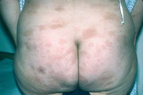
Mycosis Fungoides (Cutaneous T-Cell Lymphoma)
“Smudgy” patches and plaques or tumors are noted, usually on the buttocks and trunk.
Lesions are often pruritic.
A biopsy may demonstrate a cutaneous T-cell lymphoma.
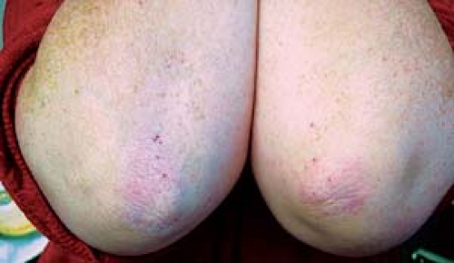 3.9 Eczema (lichen simplex chronicus). Note the lichenification and poor demarcation of these lesions (they blend gradually into normal surrounding skin). |
 3.10 Nummular eczema. Pruritic, round, nummular (coin-shaped), itchy patches with erythema, crusts, and lichenification. |
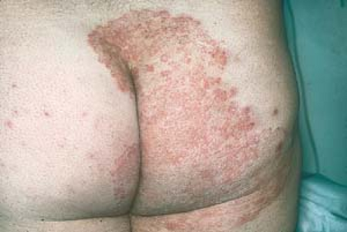 3.11 Tinea corporis. Asymmetric erythema and KOH-positive scale (“active border”). This patient has a “psoriasiform” plaque. |
General Principles
Because psoriasis is a chronic skin condition, any approach to its treatment must be considered for the long term. Treatment regimens must be individualized according to age, sex, occupation, personal motivation, other health conditions, and available resources. Disease severity is defined by the number and extent of plaques present, as well as by the patient’s perception and acceptance of the disease. Treatment, therefore, must be designed with the patient’s specific expectations in mind.
Therapy for psoriasis is aimed at decreasing size and thickness of plaques, reducing pruritus, alleviating arthritic symptoms if present, and improving emotional well-being. Ultimately, the determination of successful treatment includes both objective and subjective measures.
The types of treatment selected are determined by some of the following factors:
Age of the patient: Many oral agents that are used to treat severe psoriasis, such as methotrexate and oral retinoids, are less likely to be used in children. Very young children, particularly infants, are not able to cooperate with phototherapy treatment (described later).
Type of psoriasis.
Site and extent of involvement.
Health care provider’s experience in managing psoriasis. (Management of mild to relatively moderate psoriasis can be performed by primary care clinicians. Moderate to severe psoriasis is best treated by dermatologists.)
Availability of facilities, such as a phototherapy unit.
Presence of psychosocial problems, such as anxiety, depression, alcoholism, and substance abuse.
Three basic treatment modalities are available for the overall management of psoriasis: (a) topical agents, (b) phototherapy, and (c) systemic agents, including biologic therapies. These treatments may be used alone or in combination.
Topical therapy is the first-line approach in the treatment of plaque psoriasis (Tables 3.1 and 3.2). A number of topical treatments are available (e.g., corticosteroids, coal tar, anthralin, calcipotriene, tazarotene). No single topical agent is ideal, and many are often used concurrently in a combined approach. Auxiliary agents such as scale-removing keratolytics can often be added to these preparations.
Phototherapy and systemic therapy are initiated only after topical treatments have been unsuccessful (see also the later section titled “Generalized Plaque Psoriasis”). These treatments are considered for patients with very active psoriasis or patients who have disease that is physically, psychologically, or socially disabling.
Specific Treatment of Localized Plaque Psoriasis
Topical Corticosteroids
The use of a potent topical steroid for a limited period, followed by a less potent topical steroid for maintenance, is the most common method for treating psoriasis and many inflammatory dermatoses (see “Introduction”).
Advantage
Rapid onset in decreasing erythema, inflammation, and itching
Disadvantages
Drug tolerance (tachyphylaxis) is not uncommon
Expensive and time consuming, especially when large areas are treated
Occlusion of Topical Steroids
Generally a medium- or high-potency agent is applied and then covered with polyethylene wrap (e.g., Saran Wrap) for several hours or overnight, if tolerated. Cordran tape (see “Introduction: Topical Therapy”) is similarly effective (see also Chapter 2, “Eczema”).
Advantage
Increases the absorption, and thus potency, of topical steroids
Table 3.1 Short List of Topical Steroids | ||||||||||||||||||||||||||||||||||||||||||
|---|---|---|---|---|---|---|---|---|---|---|---|---|---|---|---|---|---|---|---|---|---|---|---|---|---|---|---|---|---|---|---|---|---|---|---|---|---|---|---|---|---|---|
| ||||||||||||||||||||||||||||||||||||||||||
Table 3.2 Other Topical Agents
Stay updated, free articles. Join our Telegram channel
Full access? Get Clinical Tree
 Get Clinical Tree app for offline access

|
|---|
