Fig. 39.1
Clinical photo of two adjacent foci of PH from the leg of a young male
Pathology
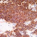
PH is characterized by an infiltrative proliferation of spindle cells arranged in sheets and fascicles (Figs. 39.2 and 39.3). In some areas, the cells exhibit an epithelioid morphology, with abundant cytoplasm and ill-defined cell borders. Some authors have compared the neoplastic cells to rhabdomyoblasts, due to their glassy and brightly eosinophilic cytoplasm (hence, the name “pseudomyogenic” hemangioendothelioma) (Fig. 39.4). Most cases show mild to moderate cytologic atypia, with few mitoses (<5 per 50 HPF). Other reported features include papillomatous epidermal hyperplasia, ulceration, and increased stromal neutrophils (Fig. 39.5). By immunohistochemistry, the cells are positive for pancytokeratin, CD31, FLI-1, and INI-1 (hSNF5/SMARCB1) (nuclear INI-1 expression retained). Two recent studies have also reported positivity for ERG, further supporting the tumor’s presumed vascular origin. CD34, S-100 protein, and desmin are negative. Recently, 3 tumors from a single patient were found to harbor a balanced t(7;19)(q22;q13) translocation. In a study of 9 additional cases, an unbalanced der(7)t(1;19) translocation was detected in one case. Further characterization of this genetic anomaly may lead to improved diagnostic accuracy.
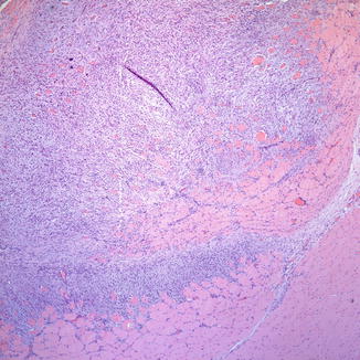
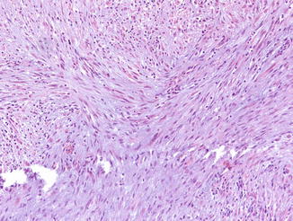
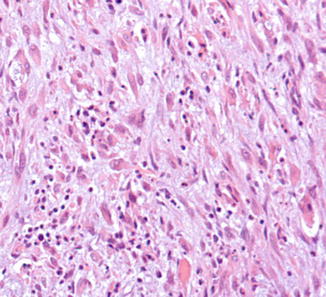
Fig. 39.2
Neoplastic proliferation of spindle cells arranged in sheets and dissecting through the muscle
Fig. 39.3
Intersecting fascicles of spindle cells
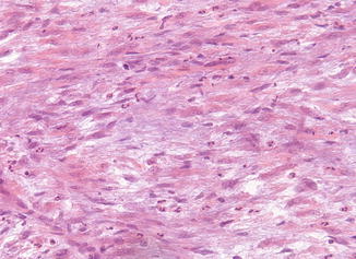
Fig. 39.4
In some areas, the neoplastic cells contain abundant, eosinophilic cytoplasm imparting a rhabdomyoblast-like appearance