Fig. 26.1
The patient presents with a gradual chronic swelling and thickening of the upper eyelid
Pathology
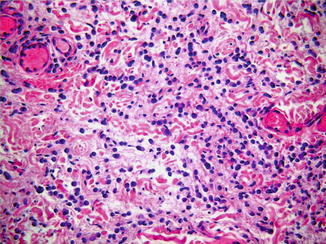
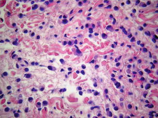
Infiltrating cords of rectangular, cuboidal, or polyhedral tumor cells forming “single-file pattern” are present throughout the dermis (Fig. 26.2). Loosely cohesive infiltrating sheets are also noted. Most of the tumor cells have an abundant eosinophilic or amphophilic cytoplasm with intracytoplasmic vacuoles resembling histiocytes (Fig. 26.3). A small number of cells exhibit a signet-ring appearance (Fig. 26.4). The nuclei are round with finely granular nuclear chromatin and distinct nucleoli. Mitoses can be found. Periodic acid–Schiff positive highlights intracytoplasmic vacuoles. There is little or absent gland formation. The tumor cells showed positive immunoreactivity to a panel of cytokeratins (CAM 5.2, CK7, AE1/3, 34BE12, and MNF116), gross cystic disease fluid protein-15 (GCDFP-15), carcinoembryonic antigen (CEA), epithelial membrane antigen (EMA), CD15, and Ber-EP4 (Fig. 26.5). Staining for CK20, CDX-2, p63, D2-40, and estrogen receptor (ER) is negative. However, some cases in the literature are stained positively by estrogen receptors and progesterone receptors.


Fig. 26.2
Infiltrating cords of rectangular, cuboidal, or polyhedral tumor cells forming “single-file pattern” are present throughout the dermis. There is little or absent gland formation Courtesy C.Tomasini,Turin,Italy

Fig. 26.3
Most of the tumor cells have an abundant amphophilic cytoplasm
Stay updated, free articles. Join our Telegram channel

Full access? Get Clinical Tree


