Fig. 45.1
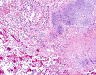
PC-EO displays sheets of hyperchromatic spindle cells with an intervening network of densely eosinophilic osteoid which shows focal calcification

Fig. 45.2
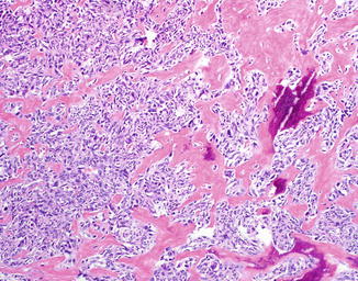
In addition to osteoid production, cartilaginous differentiation may also be seen in PC-EO

Fig. 45.3
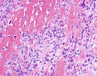
Markedly atypical spindle cells intimately associated with strands of densely eosinophilic osteoid are the hallmark of PC-EO. The presence of purple/basophilic calcification is useful in confirming the presence of true osteoid

Fig. 45.4
Atypical tumor cells are often entrapped within the osteoid in PC-EO

Fig. 45.5
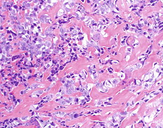
Atypical tumor cells are often entrapped within the osteoid in PC-EO. Strands of osteoid are seen between individual tumor cells
Differential Diagnosis
The most important differential diagnoses to exclude are cutaneous metastasis of osteosarcoma and cutaneous extension of a deep osteosarcoma of bone or deep soft tissue; both require clinical and radiographic correlation. Matrix producing melanoma may closely mimic extraskeletal osteosarcoma, and osteosarcomatous transformation can rarely be seen in sarcomatoid carcinoma, but both of these possibilities can typically be excluded by immunohistochemistry. Other benign and malignant skin tumors and reactive processes may sometimes display metaplastic ossification, a reactive phenomenon which may lead to confusion with osteosarcoma. Unlike the fine strands of osteoid intimately associated with malignant tumor cells that typify osteosarcoma, metaplastic ossification is usually a well-differentiated osteoid similar to mature bone. Metaplastic ossification is a typical feature of myositis ossificans, ossifying fibromyxoid tumor of soft parts, and osteoma cutis. It can also be seen as a secondary finding in a wide variety of benign and malignant neoplasms including pilomatricoma, intradermal nevus, desmoplastic melanoma, basal cell carcinoma, dermatofibroma, chondroid syringoma (mixed tumor), melanoma, and other sarcomas.
Stay updated, free articles. Join our Telegram channel

Full access? Get Clinical Tree


