Introduction
Venous disease encompasses a wide spectrum of clinical manifestations, from asymptomatic spider veins on the legs, to intermittently bulging branches of the greater saphenous vein extending across the knee, to dull achy pain in the posterior calf after prolonged standing. The science of treatment of venous disease, phlebology, has roots dating to the ancient Greeks in 400 BC, at which time venous disease was recognized as undesirable and unsightly. Procedures involving the use of instrumentation to traumatize veins were described by Hippocrates in the 4th century BC and procedures such as vein stripping were routinely practiced in the years to follow.
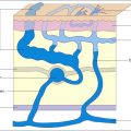
In modern days, venous disease still presents a formidable challenge to diagnose and treat. Venous insufficiency, which is caused by valvular incompetence in the deep or superficial venous system, is the most common form of venous disease. Venous disease affects 40–55% of the population, with common symptoms of leg pain, swelling, and skin changes. Superficial venous insufficiency occurs when a high-pressure leakage develops between the deep and superficial systems, or within the superficial system itself, followed by sequential failure of the venous valves in superficial veins. The two major divisions of the superficial system are the great saphenous vein (GSV) and small saphenous vein (SSV). Venous insufficiency in this system allows venous blood to escape from its normal flow path and to flow in a retrograde direction down into an already congested leg. Over time, incompetent superficial veins acquire the typical dilated and tortuous appearance of varicosities. Furthermore, insufficiency can lead to chronic morbidity in the form of ulcerative and edematous skin changes in the lower extremities.
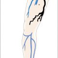
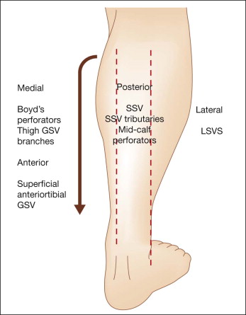
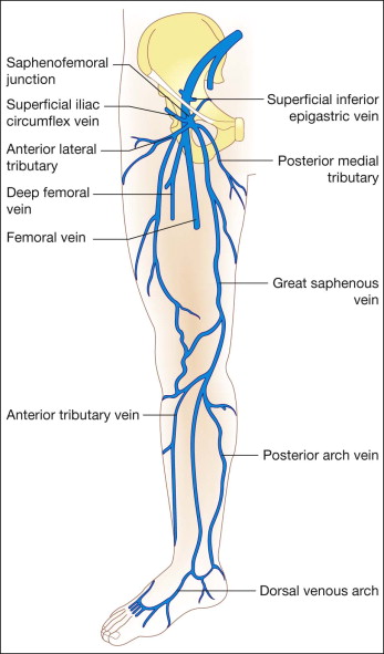
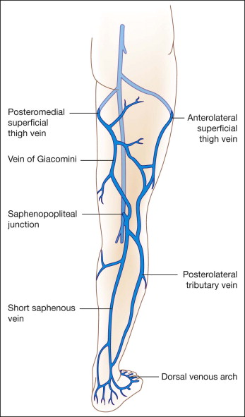
Further ‘downstream,’ changes involving the smaller branching vessels such as unsightly or symptomatic venulectasis and/or telangiectasias, are a major consequence of superficial venous valvular insufficiency. To optimize treatment of varicosities and telangiectasias, pattern recognition of common clinical manifestations is highly recommended. For a regional consideration of the anatomy that gives rise to varices, it is helpful to divide the thigh and calf into eight quadrants: lateral, medial, anterior and posterior ( Figs 2.1 , 2.2 ). A regional approach can lead to some unavoidable repetition because many veins extend through many regions or have tributaries that cross many boundaries. Clinical photographs accompanied by simplified diagrams are helpful for identification of the root causes of an unfamiliar pattern of reflux. An understanding of normal venous anatomy is essential for a thorough understanding of venous disease ( Figs 2.3 , 2.4 ). Knowledge of the location of the major perforator veins, which connect the deep venous system to the superficial system, is also important for determining the etiology of patterns of varicosities ( Fig. 2.5 ).

Treatment Approach
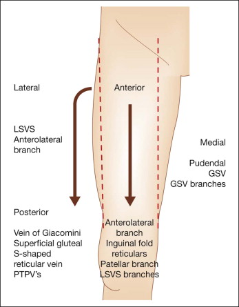
It is best to approach the clinical examination of venous disease by visualizing the lower extremities from superior to inferior, starting from the upper thigh to the calf, and concluding with the ankle. Above and below the knee, a division of four compartments can be made in the anterior and posterior planes. When considering the thigh, the presence of reflux in the GSV can manifest in the appearance of varicosities in the medial thigh compartment ( Box 2.1 ).
| Medial thigh | |
| Incompetent sapheno-femoral junction | Hidden reflux at saphenofemoral junction (only palpable on standing) |
| Superficial axial branch veins (medial tributaries) of GSV | Resistant telangiectatic matting (just above knee) due to GSV reflux |
| Distal saccular saphenous vein dilation (just above knee) | |
| Pudendal vein | Mid-thigh perforators |
| Posterior thigh | |
| Superficial gluteal | S-shaped reticular vein of posterior thigh |
| Posterior thigh perforators emptying into LSVS | S-shaped reticular vein of posterior thigh |
| Vein of Giacomini | |
| Lateral thigh | |
| Lateral subdermic venous system (Albanese) – most common cosmetic pattern | Anterolateral tributary of the GSV |
| Branch varicosity of small saphenous vein | |
| Anterior thigh | |
| Antero-lateral tributary of GSV | |
| Inguinal fold reticulars | Incompetent sapheno-femoral junction |
| Superficial axial branch veins (lateral tributaries) of GSV | Small anterior branches of the LSVS |
| Patellar |
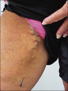
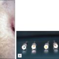
Pudendal veins are 3–4 mm blue reticular varicosities that can be seen extending from the external genitalia. These indicate reflux in the pudendal tributary of the GSV and when they become engorged, as may happen during sexual activity or menses, pain can occur. Treatment of these varicosities by sclerotherapy is easily accomplished in the absence of saphenofemoral junction incompetence.
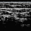
Because the GSV typically lies deep and is surrounded by fascial layers, it may be difficult to clinically appreciate prominence that accompanies reflux in this anatomic compartment. In the case of saphenofemoral junction reflux, duplex ultrasound (DUS) is usually necessary to confirm reflux. Prolonged standing may facilitate visualization of the GSV ( Fig. 2.6 ). As the GSV courses distally, superficial tributaries can become varicose as they accept reflux from above and below. In the most distal aspect of the thigh medially, just above the knee, the GSV can emerge from the fascial layers and may become apparent as an enlarged bulbous segment ( Fig. 2.7 ). Clinical manifestations of a refluxing GSV can vary based upon the degree of reflux and branch involvement ( Fig. 2.8 ).