(1)
Anatomic Pathology Laboratory, “Oncoteam Diagnostic”, Monza Hospital, Bucharest, Romania
(2)
Department of Pathology, University of Arkansas for Medical Sciences, Little Rock, AR, USA
(3)
Department of Dermatology, “Dr. Carol Davila” Central University Emergency Military Hospital, Bucharest, Romania
Keywords
Epidermotropic eccrine carcinomaMalignant hidroacanthoma simplexMalignant intraepidermal eccrine poromaSweat gland carcinomaDysplastic poromaEccrine porocarcinomaPoroepitheliomaMalignant eccrine poromaMalignant syringoacanthomaIntroduction
First described by Pinkus and Mehregan in 1963 as epidermotropic eccrine carcinoma, porocarcinoma (PC), the malignant counterpart of poroma, represents a very rare tumor derived from the dermal sweat gland duct and the acrosyringium.
Clinical Features
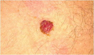
The tumor typically appears in the elderly, in the fifth to seventh decades of life. The average age at presentation is 69 years (range 8–91). Men and women are equally affected. The most common location is the lower extremity. Other common sites are the head and neck region, the trunk and the upper extremity. Occasionally, the tumor may appear on the genitals, lower and upper eyelid, ear, and the lip. The time interval between the appearance of the tumor and the diagnosis varies, but is rather large and ranges from several months to 50 years. Although most of the lesions appear de novo, they can also develop in a preexisting poroma or hidroacanthoma simplex in a significant proportion of cases. There are isolated reports of porocarcinoma arising in nevus sebaceus, linear epidermal nevus and seborrheic keratosis. The lesion may present as a slowly or rapidly growing nodule, a verrucous plaque, an ulcerated vegetating tumor, a pedunculated growth, or as a partly erosive multinodular plaque. It can have variable colors (erythematous, violaceous, brownish, or skin colored) and sizes (range 1–20 cm). Given its protean morphology, porocarcinoma can be mistaken for both benign and malignant neoplasms, such as a verruca vulgaris, seborrheic keratosis, poroma, pyogenic granuloma, angioma, basal cell carcinoma, Bowen’s disease and squamous cell carcinoma, melanoma, atypical fibroxanthoma, and a cutaneous metastasis from visceral carcinoma (Fig. 25.1).


Fig. 25.1
Porocarcinoma. An exophytic reddish tumor on the anterior trunk of a 65-year-old man. It can easily be mistaken for a vascular tumor or an amelanotic melanoma (Courtesy of Cristian Tiglea, MD)
Pathology
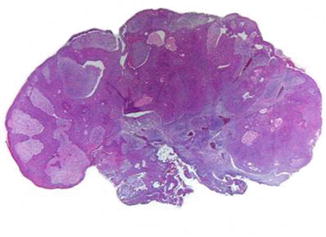
At scanning magnification porocarcinoma may exhibit some similarities to a poroma such as the presence of anastomosing epithelial downgrowths with multiple foci of attachment to the epidermis. However, the tumor has a silhouette of a malignant neoplasm, being large, asymmetrical, and poorly circumscribed and usually extending throughout the dermis and subcutaneous fat (Fig. 25.2). Some neoplasms have sharply delineated margins and a broad, pushing lower border, while others manifest an infiltrative growth pattern (Figs. 25.2 and 25.3). At higher magnification, the neoplasm exhibits two types of cells, basophilic poroid and cuticular with eosinophilic cytoplasm. Signs of ductal differentiation in the form of intracytoplasmic vacuoles and/or well-formed ducts lined by an eosinophilic cuticle are usually seen (Fig. 25.4). The cells have crowded, pleomorphic, and hyperchromatic nuclei, with many abnormal mitotic figures (Fig. 25.5). Intraepidermal spreading of tumoral cells can be seen in primary porocarcinomas, (Fig. 25.6) but is more prominent in metastatic lesions. Purely in situ cases also exist. Variants with clear cells, spindle cells, giant cells, and mucinous cells can sometimes be encountered. Some tumors may display colonization by dendritic melanocytes and melanin pigment accumulation into the cells. Areas with bowenoid changes may be found mainly in the superficial aspects of the neoplasms. Extensive squamous differentiation in the form of dyskeratotic cells and keratin whorls is a rare finding within the invasive component of the lesion. The neoplasm is embedded in a myxoid, vascular, or desmoplastic stroma. Comedo-like necrosis and cystic degeneration are other encountered features. Identification of lymphovascular invasion or perineural infiltration signifies a worse prognosis.


