Figure 17.1
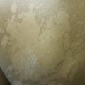
Ill-defined, hypopigmented slightly scaly patches on the right cheek
Clinical Differential Diagnosis
The findings were thought to be most consistent with pityriasis alba. The differential diagnosis for pityriasis alba includes tinea versicolor, nevus depigmentosus, vitiligo, nummular dermatitis, hypopigmented mycosis fungoides, tuberous sclerosis, and leprosy (Table 17.1).
Table 17.1
Differential diagnosis
Diagnosis | Distinguishing features |
|---|---|
Tinea versicolor | More extensive with more evident scale |
Common location: on trunk | |
Nevus depigmentosus | Usually a persistent, solitary, and hypopigmented patch with scalloped borders |
Present at birth or before age 3 | |
Common location: on trunk | |
Vitiligo | Completely depigmented and very well demarcated macules/patches |
Accentuated by Wood’s lamp | |
Nummular dermatitis | Usually more well-defined, raised plaques with more prominent scale |
Associated with intense pruritus | |
Hypopigmented mycosis fungoides | Rare condition |
Common location: on trunk | |
Tuberous sclerosis | Present at or around birth |
Associated with other systemic manifestations | |
Common location: on trunk | |
Leprosy | May have associated anesthesia/hypoesthesia with nerve involvement on histology |
Histopathology
A biopsy was not performed in this case because the physical examination was felt to be diagnostic. Pityriasis alba (PA) is typically nonspecific on histopathology, with early lesions demonstrating spongiosis, follicular plugging, focal parakeratosis and acanthosis, atrophic sebaceous glands, superficial perivascular lymphocytic infiltrate, and dermal edema. Late lesions show nonspecific changes of hyperkeratosis, focal parakeratosis, and spongiosis. There is a variable reduction of melanin in the basal layer but no significant difference in melanocyte count (In et al. 2009).
Diagnosis
Pityriasis Alba
Case Treatment
The nature and course of this condition was discussed with the patient as well as treatment options, including monitoring and topical therapies. Hydrocortisone 1 % cream daily 1 week on, 1 week off as needed for pruritus was prescribed. In addition, the importance of dry skin care, including frequent emollient use, and sun protection was emphasized.
Discussion
Pityriasis alba (PA) is a common skin disorder in all skin types and ethnicities (In et al. 2009). It is characterized by asymptomatic or mildly pruritic hypopigmented ill-defined irregular patches covered with fine scale located primarily on the face but can also involve the trunk and limbs when diffusely involved. Epidemiologic studies have demonstrated that PA is the most common hypopigmentary disorder in children. It most frequently affects children 3–16 years of age but may occur in adulthood as well (In et al. 2009). It has a higher prevalence in individuals with darker skin (FST III-VI), with one study demonstrating 98.1 % of PA patients within these skin types (Blessmann Weber et al. 2002). This may be because the lesions are more apparent in darker skin types who tan more easily except in the areas with PA lesions (Blessmann Weber et al. 2002). In addition, lesions may be more cosmetically bothersome to patients with darker complexions, making them more likely to seek treatment.
Related posts:

Stay updated, free articles. Join our Telegram channel

Full access? Get Clinical Tree


