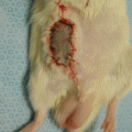
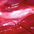
Fig. 63.1
Marking the incision site at posterior thigh of the right hindlimb of the rat
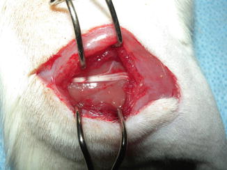
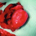
Fig. 63.2
Sciatic nerve exposure after splitting gluteal muscles

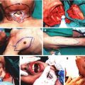
Fig. 63.3
Application of crushing force using 5-in. hemostat
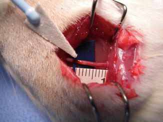
Fig. 63.4
The arrow indicates the crushed segment of sciatic nerve after removal of hemostat
Nerve Recovery Evaluation Methods
Clinical Evaluation
Functional Assessment
The animals are tested by the pinprick (PP) and toe-spread (TS) tests biweekly or weekly after nerve injury. The PP is used for evaluation of sensorial recovery. A pinching stimulus using simple standardized forceps with teeth applied to skin of the hind limb of the rat from the toe to the knee joint level, until a withdrawal from the painful stimulus is elicited. The return of sensation is scored between 0 and 3 according to the response of the animal (0 = no response, 1 = response above the ankle, 2 = distal to ankle, 3 = metatarsal region). The TS is used for evaluation of motor recovery after nerve injury. The animals are held by their tails, and their toe-spread movements are graded between 0 and 3 (0 = no toe movement, 1 = any movement, 2 = toe abduction, 3 = abduction with extension).
Electrophysiologic Test
Electrophysiologic assessment is performed by somatosensory-evoked potentials evaluation (SSEP). Under ketamine anesthesia, the stimulating electrodes were placed subcutaneously into the dorsum of the foot and the Achilles tendon, ground electrode was inserted subcutaneously to the gluteal area. Then, approxiametely 2 cm sagittal incision is made on the scalp, the cranium is exposed by subperiosteal dissection. Two burr holes are created in the parietal bones of the skull and recording electrodes are placed to the epidural plane over the somatosensory cortex region. After 300 trials, the averages are obtained. SSEP measurements were done and positive (P) and negative (N) potentials of the waveform morphology are obtained. The P1 (first positive wave) and N2 (the second negative wave) are the most robust and consistent potentials, therefore they are used for comparison of sensory recovery [21]. Wave amplitudes are also measured for assessment of axonal regeneration. Latencies and amplitude values are evaluated as a percentage from the unoperated side (P1 %, N2 %, Amp %) to eliminate the anesthesia factor.
Histomorphometric Evaluation
Gastrocnemius Muscle Weight Index (GMI)
Evaluation of the effects of denervation on the muscle is done by assessment of the GMI. The gastrocnemius muscles from both sides are carefully disected and excised at the end of the follow-up period and wet muscle weights are measured using a digital scale. The muscle weight index is calculated as a percentage of operated/nonoperated sides that represents the recovery in denervation atrophy of the muscle on the operated side. After formalin fixation, sections are taken from cross-section of muscles. Stained slides are evaluated for muscle atrophy, mean muscle fiber diameter and fiber cross-sectional area using softwares.
Nerve Morphometry and Morphology
At the end of the follow-up period, under ketamine anesthesia sciatic nerves are dissected proximally and distally from the crushed nerve segment. Sections are taken from the proximal nerve to the crushed nerve, the middle of the crushed part and distal to the crushed injury site and are fixed in 4 % glutaraldehyde for final processing. The contralateral side sciatic nerve is served as a control group. Analysis of the histomorphometric parameters, such as axon count, axon density, total number of myelinated fibers, axon diameter, fiber diameter, myelin thickness and g-ratio are calculated using software programs. The g-ratio is calculated by dividing the axon by the fiber diameter and myelin thickness is computed by a substracting the axon from the fiber diameter and then dividing by the factor 2. Expression of growth factors involved in nerve regeneration, such as nerve growth factor (NGF), laminin B, glial fibrillary acidic protein (GFAP) and vascular endothelial growth factor (VEGF) are evaluated by monoclonal antibodies and immunofluorescence technique.
References
1.
Related posts:
 Microcirculation Model for Invasive Animal Monitoring
 Composite Osseomusculocutaneous Thymus Allotransplantation Model
 In Vivo Chimera Model: Creation of Primary and Secondary Chimera
 Peripheral Nerve Surgery Models Crush Injury and Epineural Patch
 Heterotopic Vascularized Ovarian Autotransplantation Model in the Sheep
 Defect Repairs After Resections of Laryngeal Cancer, Hypopharyngeal Cancer, and Cervical Esophageal Cancer
Stay updated, free articles. Join our Telegram channel

Full access? Get Clinical Tree


