Perforator flaps, since their first description in 1989, have in many ways revolutionized reconstructive surgery. Whereas little more than a decade ago many surgeons were still hesitant to fully trust perforator flaps to be a reliable option, nowadays these flaps are often first choice. Investigators have to remain critical, however, of their advances and realize that not every reconstruction will require or benefit from a perforator flap, as previously well-established, nonperforator flaps still have their indication and can give excellent results. The most important skill in reconstructive surgery of the head and neck is not cutting the flap but assessing the defect, planning the reconstruction, and choosing wisely from the ever-increasing options available.
Perforator flaps, since their first description in 1989, have in many ways revolutionized reconstructive surgery. Whereas little more than a decade ago many surgeons were still hesitant to fully trust perforator flaps to be a reliable option, nowadays these flaps are often first choice. This early disbelief in the true value and sustainability of perforator flaps in general was nicely worded in the following section from the excellent flap book by Cormack and Lamberty in 1994: “… the deep inferior epigastric artery may be dissected free of rectus abdominis while preserving 1 or 2 perforators to an island of tissue consisting only of periumbilical skin, fat, and a patch of anterior rectus sheath. Nomenclature for this variant of the musculocutaneous flap has not been universally recognized but examples are extremely few and are probably to be regarded as more a demonstration of technical skill than a significant advance in flap construction ….”
The concept of perforator flaps has evolved over the years. The basic concept is that any larger named vessel in the body will give off smaller branches toward the skin on which flaps can be designed and raised as pedicled or free flaps. In the early period most, if not all, the attention regarding perforator flaps was focused on proving it was technically possible to actually dissect a very small blood vessel that perforated through a muscle and was able to vascularize a previously musculo-adipocutaneous flap as merely an adipocutaneous flap. Once this initial technical achievement had been mastered by a larger number of surgeons around the globe, further structure was brought into the new entity of perforator flap surgery. In essence there were 2 important steps to further popularize perforator flap surgery: on the one hand the further clarification of the clinical benefits and on the other hand developing classification systems to improve communication between surgeons.
The main clinical benefits that arose from perforator flaps are twofold. In the case of a musculocutaneous perforator there is a clear donor site benefit if the innervated muscle can be left in situ. The best examples for this benefit are preservation of rectus abdominis muscle in the deep inferior epigastric perforator flap and gluteus maximus muscle in the gluteal artery perforator flaps. The second benefit was the realization that potential flaps were now all over the body and all one had to do was to find a blood vessel to raise a flap. This concept was coined “the free style perforator flap.” With current knowledge the concept of “free style” probably is less spectacular, because all flaps can be found and named according to their source vessels.
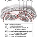
Once a critical mass of reconstructive surgeons had adopted perforator flaps, the lack of uniformity in classification of these new flaps became obvious. Several suggestions for classification schedules were proposed to facilitate and clarify communication between surgeons. Classification schedules focused around 2 thoughts, on the one hand the origin of the perforator and on the other hand the course of the perforator. At present, most communications use the source vessel to describe the flap and mention whether the course of a perforator is direct, that is, from the source vessel into the skin, or indirect, that is, from the source vessel through mostly muscle into the skin.
Source vessels for perforator flaps in the head and neck
Two major arteries are responsible for the blood supply to the head and neck area. These vessels are the common carotid, which divides into external and internal carotids, and the subclavian artery. A great number of branches stem from these vessels. Many of these branches give off their own branches or perforators that in part supply the skin of the head and neck area. Not all of these branches or perforators have clinical value as source vessels for pedicled perforator flaps. Knowledge of the location of all these vessels, however, will be of great value for safe planning and execution of tissue advancements, transpositions, and rotations in the head and neck area. Those perforator branches that have clinical potential for pedicled perforator flaps are discussed here in more detail.
The first branch from the external carotid artery is the superior thyroid artery. The only vessel from the superior thyroid artery that provides blood supply to the anterior triangle of the neck skin is the sternomastoid branch. This branch can sometimes arise directly from the external carotid artery. The next is the lingual artery, which does not give off branches to the skin. The facial artery arises immediately above the lingual artery. Over its long course from its origin in the neck until its end point near the nasal root it has several branches: submental artery, premasseteric branch, inferior labial artery, superior labial artery, lateral nasal branch, and angular branch. The occipital artery arises from the posterior surface of the external carotid artery at the level of the facial artery branch. The occipital artery gives off several perforators through the overlying muscle that supplies the skin. These perforators supply the skin in the posterior neck. The posterior auricular artery in general arises from the posterior aspect of the external carotid artery, but sometimes offshoots together with the occipital or superficial temporal artery. The auricular division of the posterior auricular artery supplies the posterior surface of the ear and the occipital division supplies the scalp posterior to the ear. The superficial temporal artery has 2 main branches, the frontal and the parietal branch. This artery has 3 main branches supplying the skin: the transverse facial artery (that sometimes arises directly from the external carotid artery), the auricular branches, and the zygomatico-orbital branches. The maxillary artery supplies skin through the infraorbital and mental branches.
The internal carotid artery supplies the skin of the face through the cutaneous branches of the ophthalmic artery, which supply the forehead through the supraorbital and supratrochlear arteries, the periorbital skin through the medial palpebral and supraorbital arteries, and part of the nasal skin through the external nasal artery. All these branches of the internal carotid artery anastomose significantly with its local counterparts from the external carotid artery.
The thyrocervical trunk of the subclavian artery gives rise to the transverse cervical artery, which has a deep and superficial branch, and the suprascapular artery. The supraclavicular artery can arise from either of these two vessels. The anatomy in this region has several exceptions with regard to the exact origin of the vessels. These vessels supply most of the skin of the neck between the clavicle and mandibular margin.
Related posts:


Stay updated, free articles. Join our Telegram channel

Full access? Get Clinical Tree


