Abstract
Fractures of the pediatric midface are usually rare and more likely to involve isolated segmental areas such as the nose or orbital–zygomatic complex depending on the age and development of the child. There is no single approach to the management of midface fractures in children and care must be individualized. The dimensions of age and growth can add to the complexity in their management. This chapter will review basic principles in their treatment.
Keywords
pediatric, midface fractures
Background
The management of pediatric facial fractures presents some unique and specific opportunities that differ from those in the adult world. It is important to recognize that children are not small adults, and when possible, treatment should be conservative. There is no single approach to the management of midface fractures in children and care must be individualized. The dimensions of age and growth can add to the complexity of their management.
The midface region traditionally involves the maxilla although the contents of this chapter will touch on regions of the orbital–zygomatic complex and nasal–orbital zone. Skeletal maturity is considered to be the age of transition to adulthood (14–16 years in females; 18–20 years in males) but it is useful to conceptualize pediatric facial trauma by the stage of dentition (primary, mixed and permanent).
Pediatric facial fractures occur with much less frequency and severity and make up between 1% and 15% of all facial fractures series. They are less common in very young children below the age of 5, representing less than 1% of all facial fractures. As children mature and start to move with increasing velocity, fracture incidence increases, representing nearly adult levels in the late teenage years. Of midfacial fractures, nasal and dentoalveolar fractures are quite common, with zygomaticomaxillary complex (ZMC) and maxillary fractures less frequently represented. When maxillary fractures do occur in young children, traditional pure Le Fort patterns are varied. Oblique fracture patterns involving the orbits and piriform aperture are more likely to occur. Nasal bone fractures and dentoalveolar fractures represent the most common facial fractures in the pediatric population. With increasing age, eruption of dentition and growth of the lower face and sinuses, there is a transition to more typical adult fracture patterns.
Pediatric facial fractures occur primarily as the result of motor vehicle collisions (MVC), falls and sports-related injuries. While changing traffic laws and safety standards (e.g. increased use of air bags, car seats, and seat belts) over the years have led to fewer MVCs, compliance varies dramatically, and up to 70% of children with MVC-related facial fractures were unrestrained at the time of injury.
Lower-velocity injuries such as falls are more common in younger children, while teenagers and young adults are more likely to suffer traffic accidents and sports-related trauma. Violent altercations are much less common causes of craniofacial trauma in children compared to adults, although this also increases with age. Child abuse as a cause of isolated maxillofacial trauma is quite rare.
Like most traumatic injuries, incidence peaks in the warmer months, attributed to more outdoor physical activity. Facial fractures are more common in boys, though this varies greatly in reports from 1.1 : 1 to 8.5 : 1 depending on the publication. This difference is less notable in younger age groups.
Surgical Anatomy
Children have a lower blood volume with overall higher cardiac output and smaller airway volume. This puts them at increased risk of complications from blood loss and airway obstruction compared to adults. As a result, the principles of initial trauma management regarding volume resuscitation and securing the airway are especially relevant in treating pediatric trauma patients. Fractures of the midface and mandible are associated with higher velocity impact and are more likely to be associated with other systemic injuries than isolated nasal or orbital fractures.
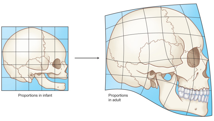
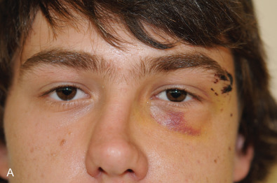
The target zone for facial trauma in the pediatric population changes with age. At birth, the cranial vault/orbital region makes up roughly an 8 : 1 ratio compared with the facial region that halves to 4 : 1 by 4 years of age and then gradually decreases to around 2.5 : 1 at skeletal maturity ( Fig. 2.4.1 ). As the facial bones are smaller in size and more retruded relative to the overlying skull at younger ages, this provides relative protection to the midface. Therefore, younger patients, especially under the age of 5, have a predilection for cranio-orbital injuries ( Fig. 2.4.2 ). As the face grows, the prominent areas (nose, zygoma, mandible) become more frequently involved and prone to injury.
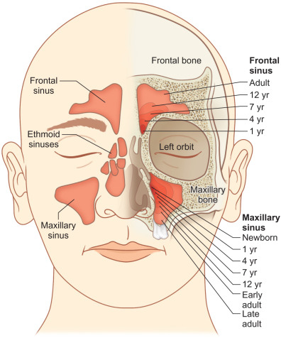
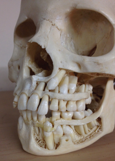
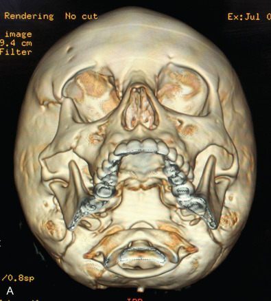
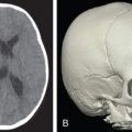
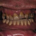
Facial fractures in children are often less displaced than those of adults and greenstick fractures are common given their more flexible bones with more mobility at suture lines and less bone mineralization. The bone exhibits a higher cancellous-to-cortical bone ratio, and without the development of the air-filled paranasal sinuses which affect the compliance of the craniofacial skeleton. At birth, the maxillary sinus is present but very small, enlarging gradually downward as the permanent dentition erupts, to fill the area previously occupied by the tooth buds. As such, it does not reach its adult size until eruption of the third molars. Ethmoid air cells are also usually present at birth, slowly expand, and are the first sinuses fully developed, which occurs around puberty ( Fig. 2.4.2 ). The sphenoid sinus first appears around age 2 and continues to enlarge until skeletal maturity, with some further septation into adulthood. The frontal sinus first appears at age 5 and continues to expand into late teen years. Increased soft tissue padding, thicker fat pads, and the strong, developing tooth buds within the maxilla and mandible also provide increased resistance to complete fractures ( Fig. 2.4.3 ). While these factors act to provide protection for the pediatric skeleton, a higher impact force per unit area is required for the facial bones to fracture in children compared with their adult counterparts and as such, there may be a higher incidence of associated injuries. The unique nature of the elastic compliant pediatric craniofacial skeleton can result in greenstick fractures and discontinuity between fracture zones ( Fig. 2.4.4 ).
Clinical Presentation
Pediatric midfacial fractures tend to occur as the result of significant trauma and usually warrant the assessment of a trauma team. A history of the injury and details of the velocity involved will give the treating physician clues as to the potential extent of the injury. Information about the use of seatbelts, helmets, height of falls, loss of consciousness, and preexisting conditions are standard elicited history. Features of the child may make obtaining a detailed history and the presence of symptoms (pain, loss of sensation, malocclusion, visual disturbances) problematic and an index of suspicion may help direct clinical investigation. It is often useful to obtain an orthodontic history given the frequency of orthodontic appliance use in children and young adults. Premorbid scans, impressions, and models may give useful information regarding the state of the preinjury dentition and occlusion.
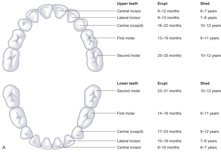
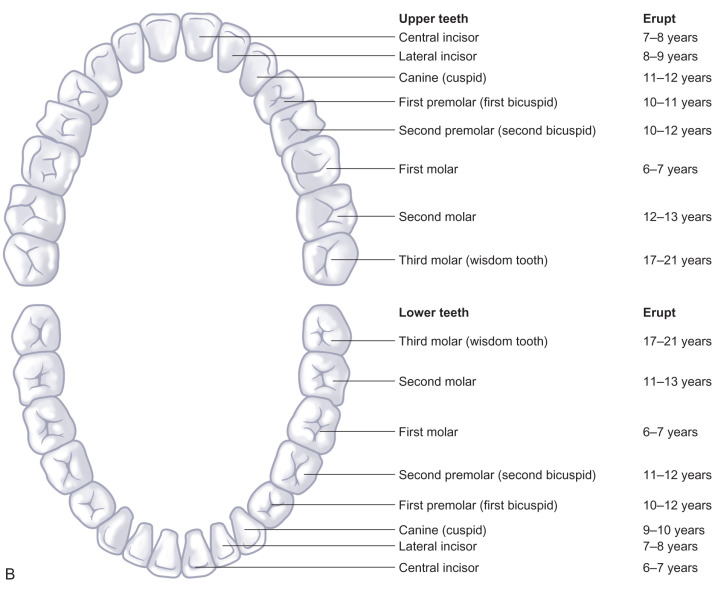
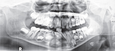
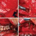
Physical examination is focused on the zone of trauma. Overlying lacerations, ecchymosis and soft tissue disruption may give clues as to directed injuries. Due to the lack of pneumatization of the sinuses and strength from unerupted tooth buds, forces applied to the midface are transferred to the dentoalveolar complex resulting in tooth avulsion and fractures. Dentoalveolar fractures are the commonest form of pediatric midface fractures and occasionally may be associated with midface gingival degloving ( Fig. 2.4.5 ). An understanding of the patterns of tooth eruption will be useful in assessing children between ages 6 to 12 in mixed dentition ( Fig. 2.4.6 ).
Systematic assessment of the integrity of the craniofacial skeleton will involve inspection for obvious asymmetries and deviations, palpation for areas of tenderness, bony steps and disruptions, and manipulation for midface instability and mobility. A careful dental examination to assess for malocclusion, mobile, lost or devitalized teeth is important. The possibility of aspirated teeth may warrant a chest X-ray.
Orbital examination is always mandated with potential midface fractures. Periorbital ecchymosis, subconjunctival hemorrhage, visual acuity, extraocular mobility, telecanthus, enophthalmos and facial sensation should be assessed. All suspected orbital injuries should have a formal ophthalmological consultation when possible. The forced duction test should be performed in any patient that cannot supply a satisfactory voluntary examination but is not tolerated in the awake child and should be done under anesthesia in order to prevent a false-positive result. A delay in diagnosis and treatment of these injuries can have significant consequences on the patient’s long-term outcome.
Midface fractures are rare in children. When present, due to the high velocities involved, associated systemic injuries may be seen in up to 88% of patients. As such, a thorough trauma and possible neurosurgical evaluation should be performed for all patients with substantial facial fractures.
Radiological Evaluation
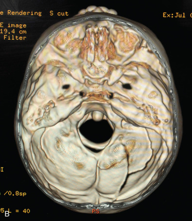
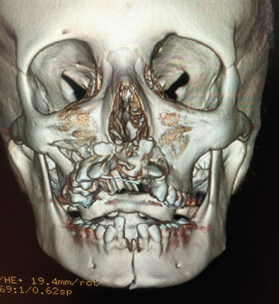
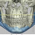
Plain films of the craniofacial skeleton, especially the midface, are of no use in the assessment of facial trauma in the pediatric population. Developing sinuses and tooth buds may obscure already overlapping planes. Computed tomography (CT) scans are necessary in order to accurately diagnose pediatric facial trauma. Fine facial cuts are necessary to adequately define facial fractures, and 3D reconstructions can be extremely useful in planning surgical intervention and teaching trainees. Cone beam CT scans and orthopantomograms (panoramic radiographs or panorex) are also useful to assess for dental injuries.
Classification
Pediatric midfacial fractures are classified anatomically and will vary according to the age of the patient. The velocity of the injury will give clues to the types of fractures seen. The orbitozygomatic and nasal regions are susceptible to lower velocity trauma than the midface maxillary complex. The degree of displacement and comminution will be used to classify these fractures. Naso-orbito-ethmoid fractures are quite rare in young children due to the flatter nasal dorsum but may be classified according to the degree of comminution of the segments containing the medial canthal ligament (Type I: large fragment; Type II: small fragment; Type III: canthal avulsion). The typical Le Fort fracture classification may be applied but usually does not occur until teen years when the maxillary sinuses have aerated and the teeth erupted. Dentoalveolar fractures are much more common in the pediatric population. Midface fractures in the pediatric age group follow oblique and unpredictable lines extending often into the cranial base and frontal areas. The palatal suture does not fuse until teen years and midline palatal fractures may be more commonly seen in children.
Surgical Indications
The indications for open reduction and rigid fixation of facial fractures in the pediatric population are more conservative than those in adults, especially in very young children. With higher metabolic activity, osteogenic potential and bone healing, children will heal more rapidly than adults, and have the added element of facial growth that may be negatively impacted by surgical intervention and periosteal stripping. Indications for surgical intervention in the midface region are both functional and aesthetic. The maxilla provides the centerpiece of the face, and the vertical and horizontal buttresses of the midface must be intact to establish appropriate facial height and width. Fracture displacement, malocclusion, severe comminution, associated soft tissue injuries and lacerations, large orbital wall defects with the potential for late enophthalmos and disruption of the medial canthal complex in NOE fractures are indications for surgical intervention in pediatric midface fractures.
Often, the compliance of the pediatric facial skeleton results in less displacement in young patients than their adult counterparts and can be treated with observation or closed reduction aided by splinting or limited MMF. General principles dictate that non- or minimally displaced fractures of the midface can be treated with observation, generally combined with a soft diet for 2–3 weeks for comfort. Displaced fractures, on the other hand, may require either closed or open reduction and fixation. If surgical intervention is necessary, quick healing times implicitly require more rapid intervention. Additionally, children require shorter periods of bony immobilization in order to heal: 2–3 weeks versus 4–6 weeks in adults.
Maxillomandibular Fixation
Maxillomandibular fixation (MMF) in children with deciduous or mixed dentition can prove challenging ( Fig. 2.4.7 ). The conical shape of the primary teeth and partially erupted permanent teeth may be difficult to adequately fixate with interdental wires and arch bars. Likewise, use of MMF screw fixation may be limited given the various permanent tooth buds present in both the maxilla and mandible. In very young children, temporary suspension wiring around the zygomatic arch or piriform aperture to the arch bar may be necessary for fixation. Fortunately, major midface fractures affecting occlusion and mandibular fractures are more common in patients with more mature dentition, such as adolescents and teenagers.
Prior to the 1960s, closed reduction with MMF was utilized for nearly all pediatric fractures. Open reduction and internal fixation (ORIF) transitioned slowly to open reduction with wires and now has become accepted for displaced fractures given technical improvements in plating technology. However, the permanent placement of hardware in growing children raises important questions in terms of cost, iatrogenic trauma to erupting tooth buds, migration of plates and effects on bony growth. Animal studies have shown slight adverse effects on growth, but no similar effects have been proven in humans. Resorbable plates may have some efficacy in pediatric fractures under the age of 10 years where a permanent implant is not desired, but do not have the shear strength of metal miniplates, and as such are not used in load-bearing bone. In general, our treatment philosophy involves consideration of hardware removal in the pediatric midface 6 months following repair in order to prevent theoretical concerns of retained hardware (bone stress shielding, bony overgrowth, dental concerns) in later years.
Surgical Techniques
Nasal Bone Fractures
Nasal bone fractures are the most common facial fractures in children. Despite this, they are often overlooked as immediate swelling and difficult compliance with intranasal exam may mask the fracture displacement. Diagnosis is made by clinical examination. Unless an NOE fracture is suspected, CT imaging is not necessary. As in adults, septal hematomas must be drained immediately to avoid septal cartilage necrosis and/or infection with potential nasal deformity (saddle nose). This is especially important in children, as a saddle nose deformity may also be associated with long-term facial growth restriction.
Displaced nasal bone fractures should be reduced within a week following injury if possible ( Fig. 2.4.8 ). Ideally this should be done in the operating room with a secure airway, and should not be attempted in the emergency department where significant epistaxis could be life-threatening. Once reduced with a Goldman elevator or Asch forceps, intranasal packing or splints for 2–4 days are helpful to maintain the reduction and to assist with hemostasis. External splinting for 7–14 days is also crucial to guide healing, but may be difficult to maintain in noncompliant younger patients. Septoplasty in the case of septal fractures is a controversial topic, and if attempted, should take care to avoid the growth center at the nasal spine of the maxilla. It is important to note that some nasal deformity may persist despite closed reduction. In the case of residual deformity outside the acute traumatic period, definitive rhinoplasty is usually deferred until skeletal maturity if possible in order to preserve nasal growth.
Related posts:


Stay updated, free articles. Join our Telegram channel

Full access? Get Clinical Tree


