Pattern analysis: Dermoscopic criteria for specific diagnoses


Dermoscopic analysis of pigmented skin lesions is based on four algorithms:


  • pattern analysis;


  • the ABCD rule;


  • Menzies’ 11-point checklist; and


  • the 7-point checklist.


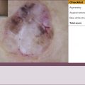
The common denominator of all these diagnostic algorithms is the identification and analysis of dermoscopic criteria found in the lesions. The majority of the dermatologists who participated in the second consensus meeting were proponents of pattern analysis. The basic principle is that pigmented skin lesions are characterized by global patterns and combinations of local criteria.


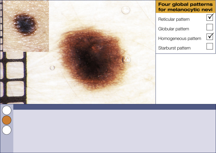
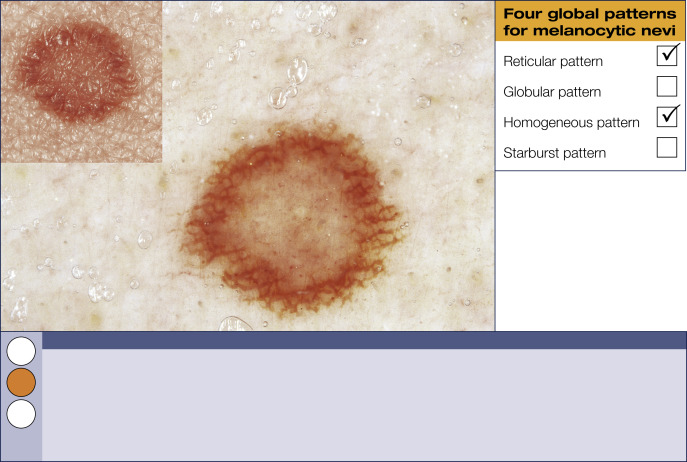
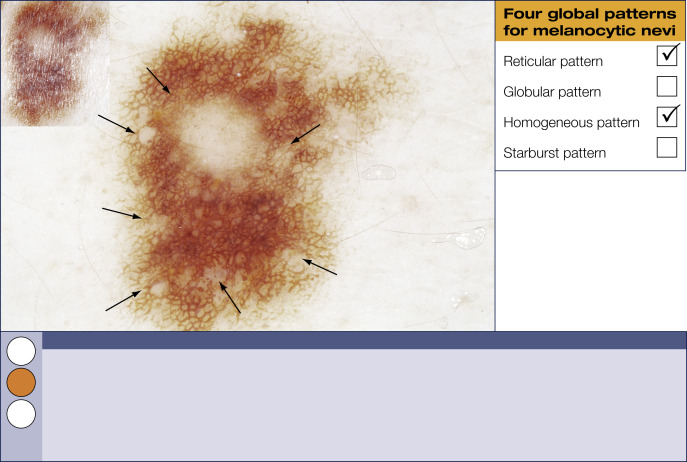
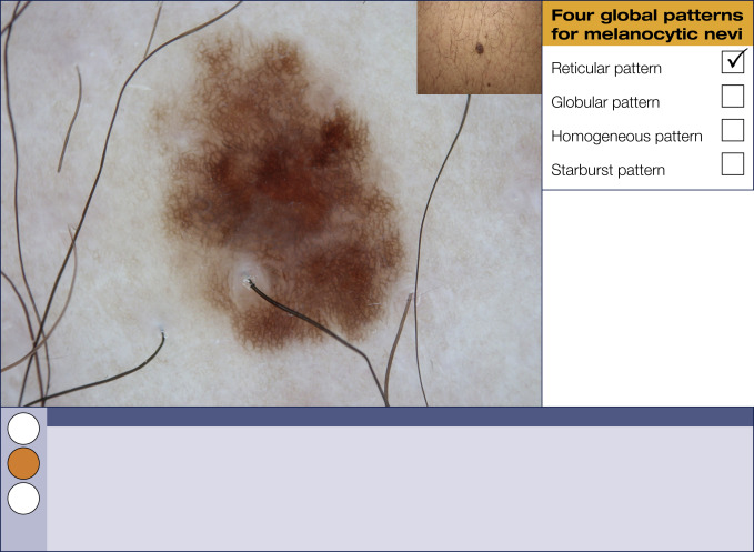
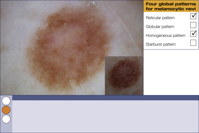
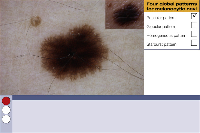
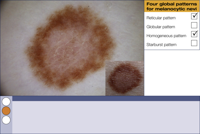
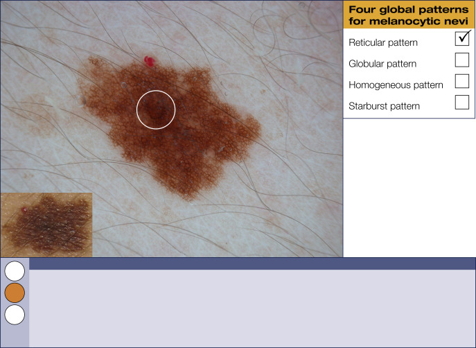
Four global dermoscopic patterns for melanocytic nevi
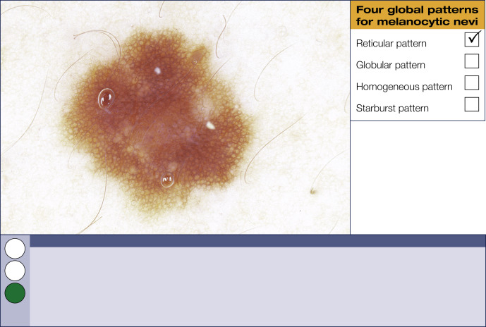
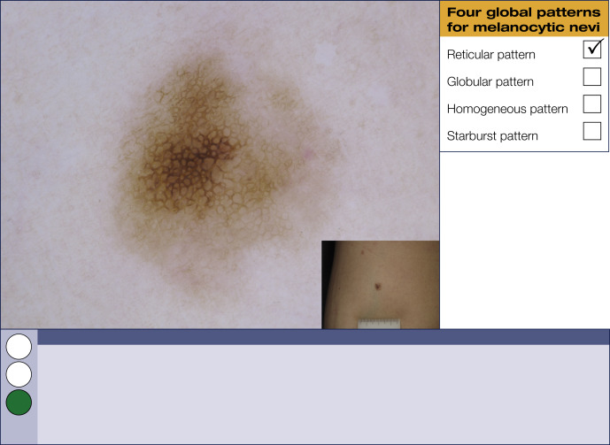
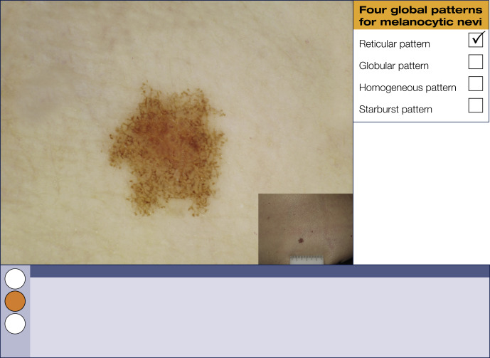
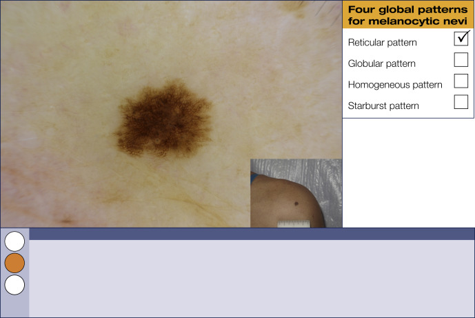
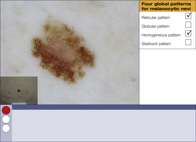
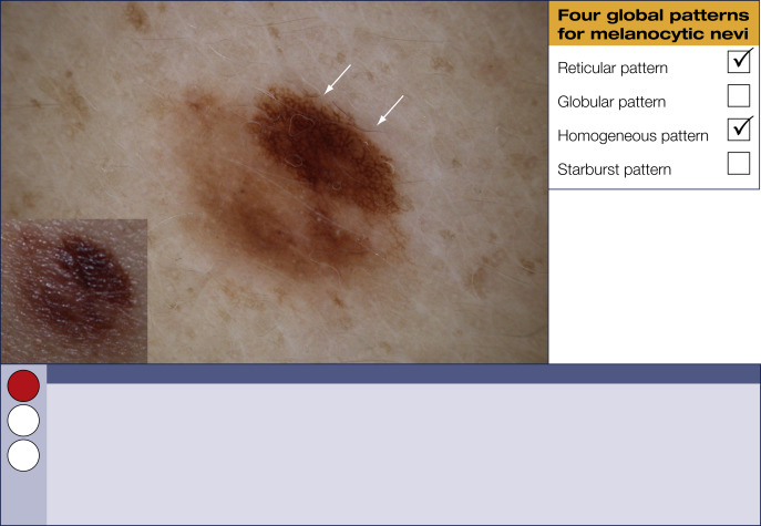
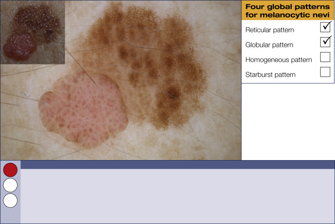
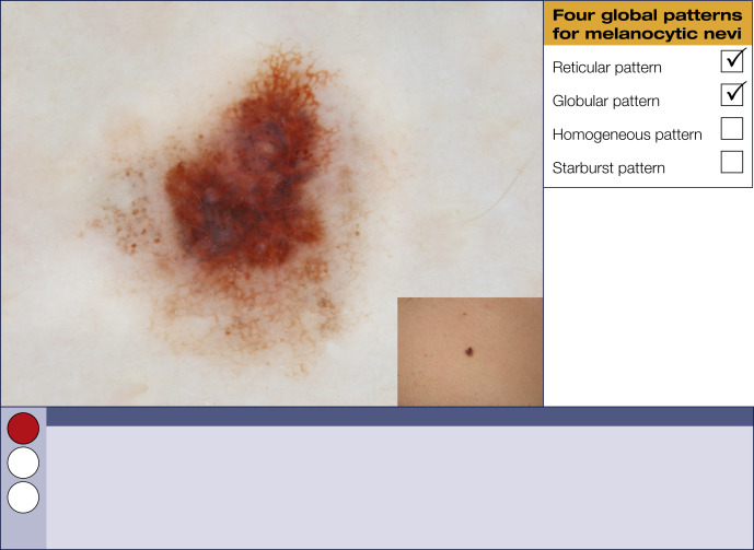
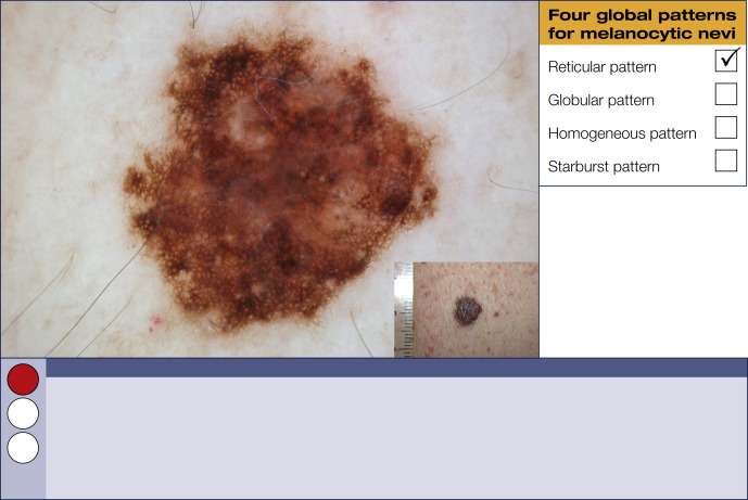
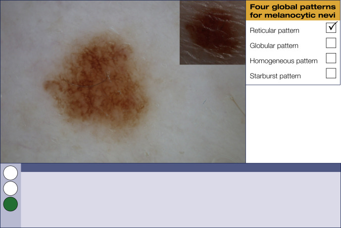
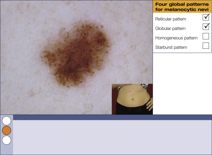
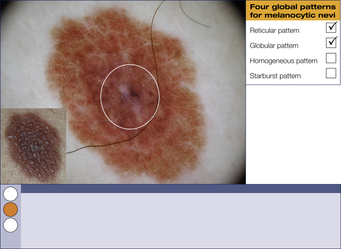
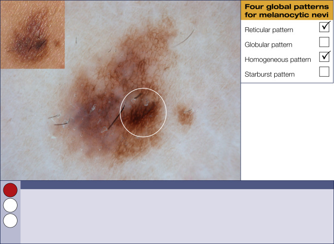
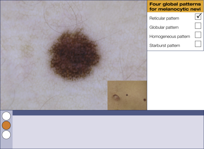
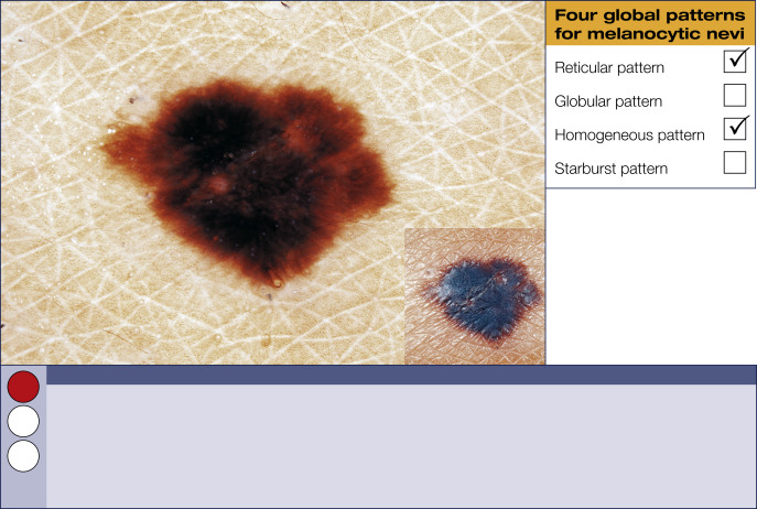
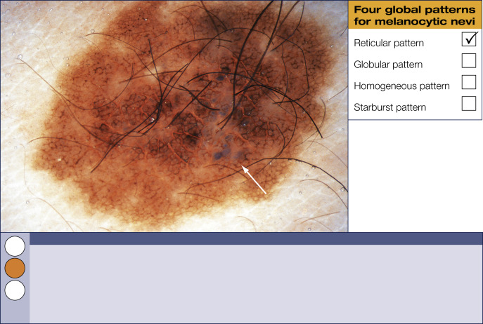
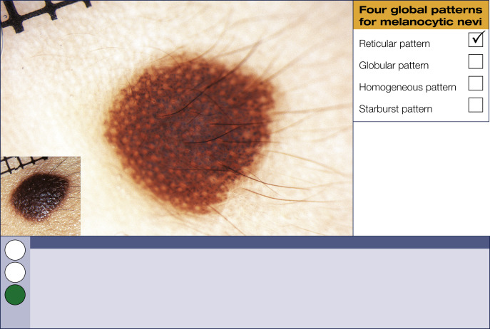
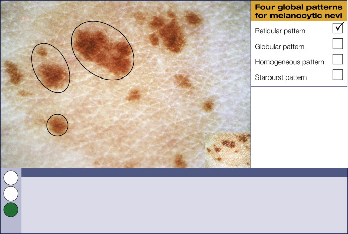
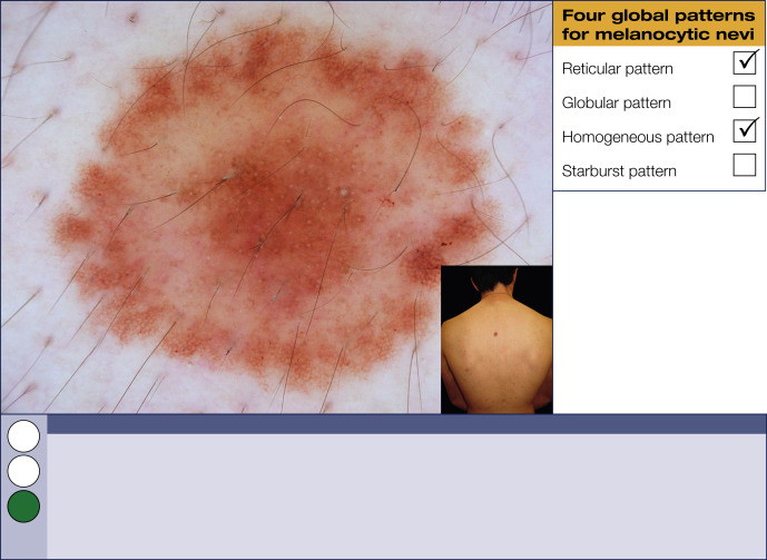
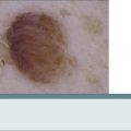

Reticular pattern


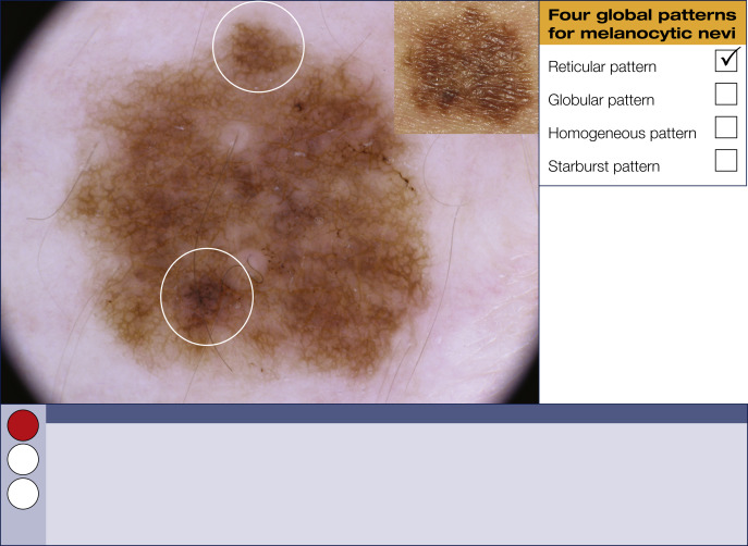
The reticular pattern is the most common global pattern in melanocytic lesions. It is characterized by a pigment network covering most parts of a lesion. The pigment network appears as a grid of line segments (honeycomb-like) in different shades of black, brown, or gray. Modifications of the pigment network vary with changes in the biologic behavior of melanocytic skin lesions, and these variations therefore merit special attention.
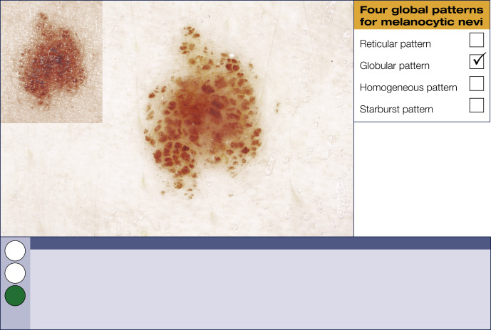
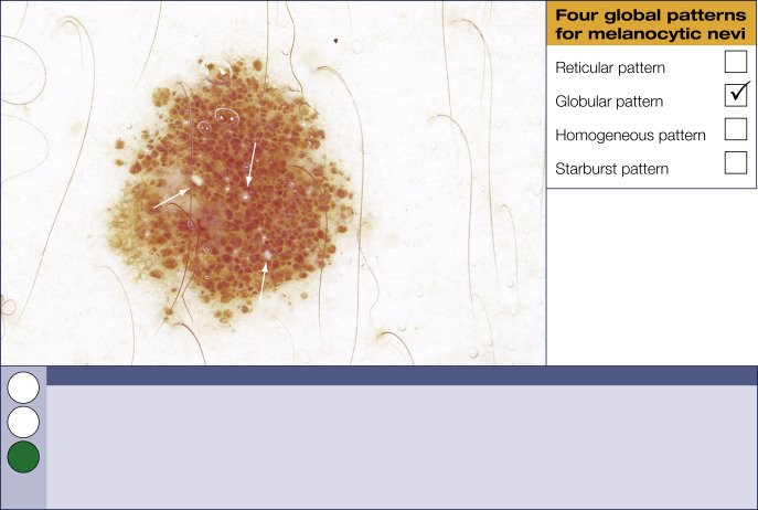
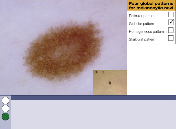
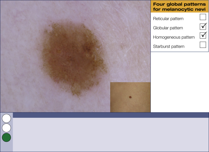
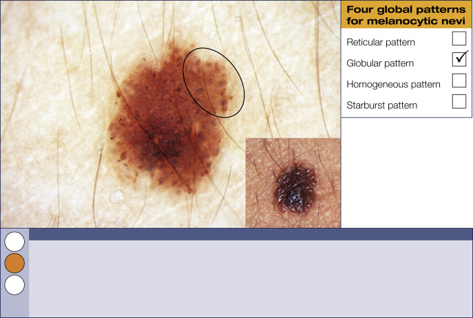
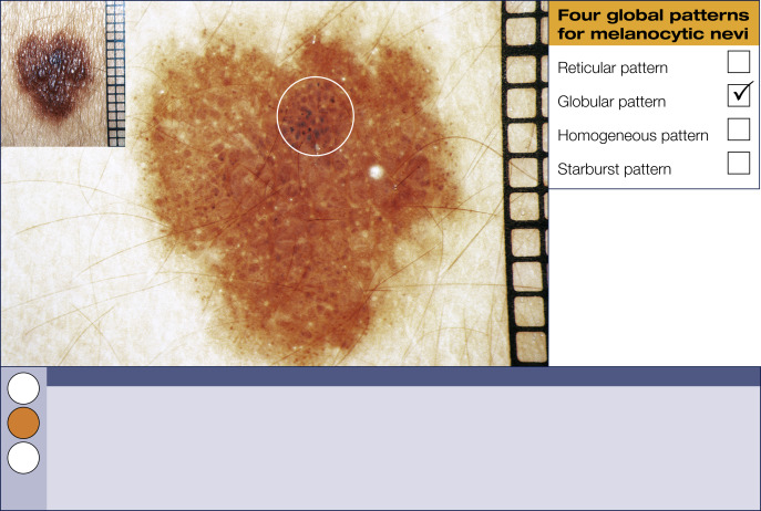
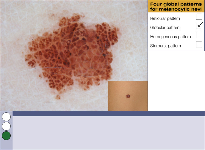
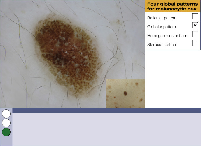
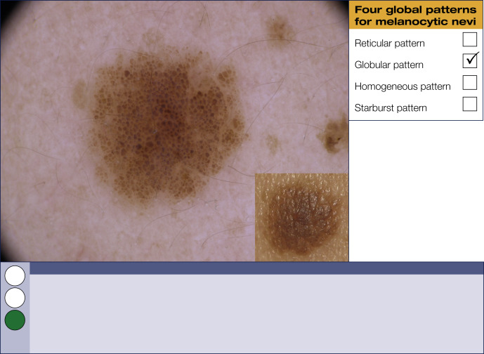
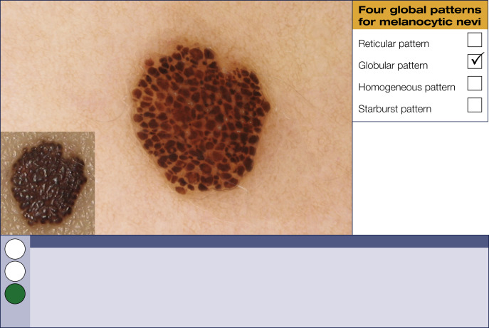
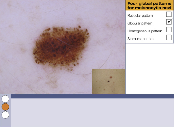
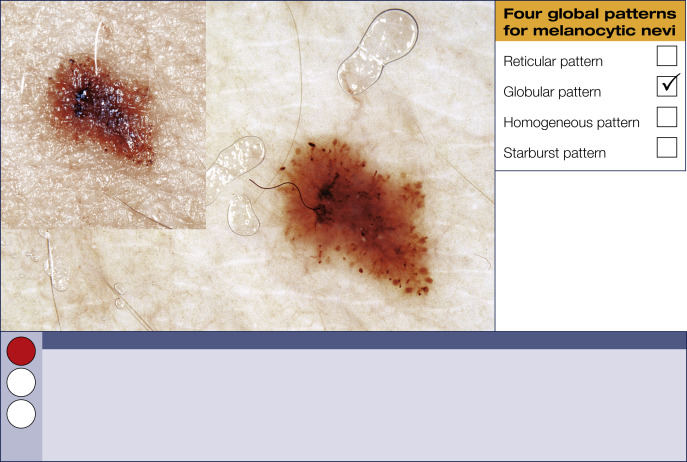
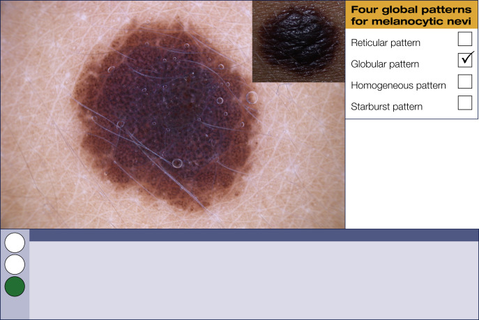
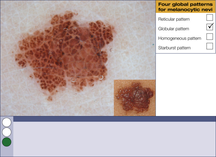
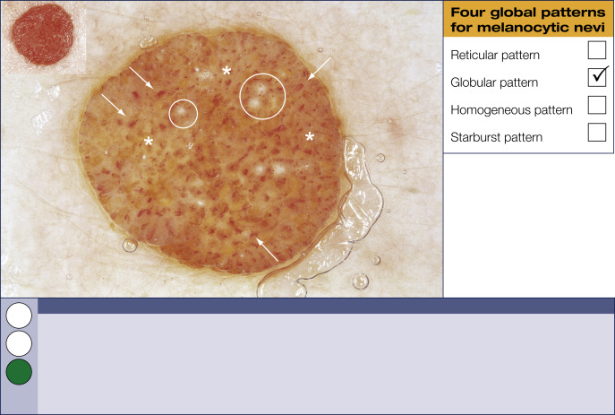
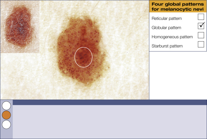
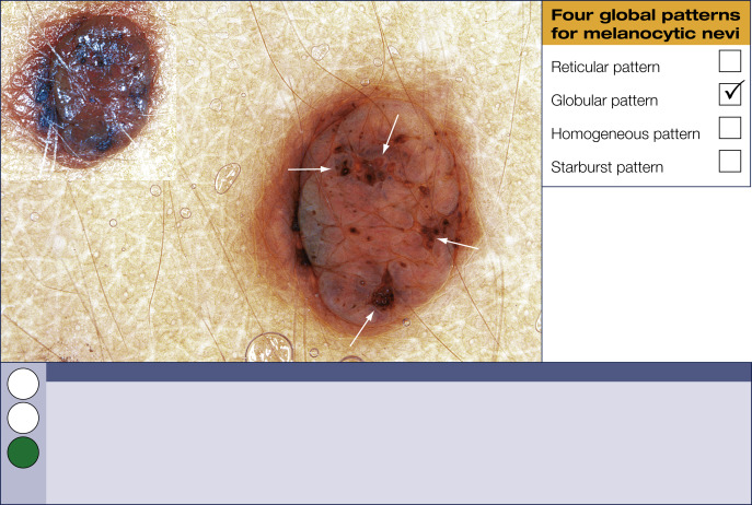
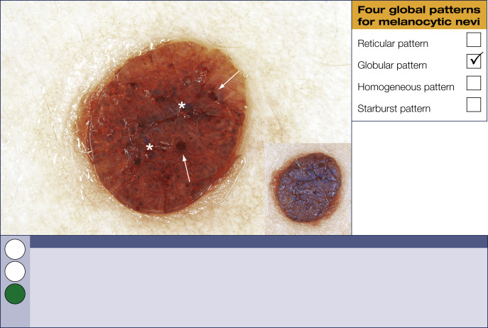
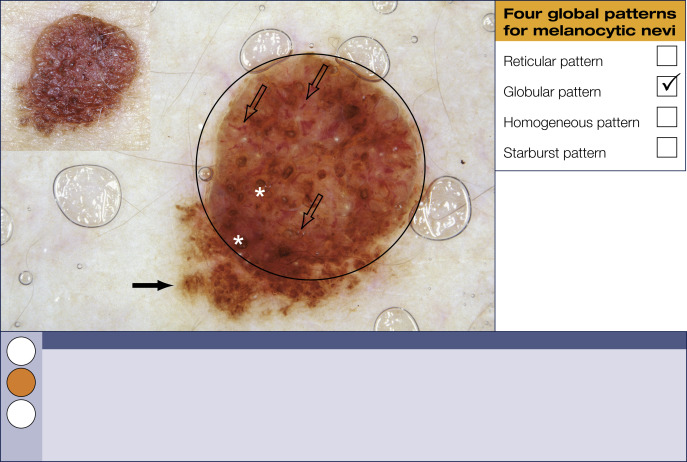
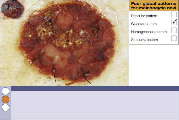
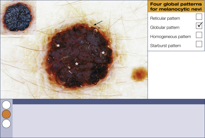
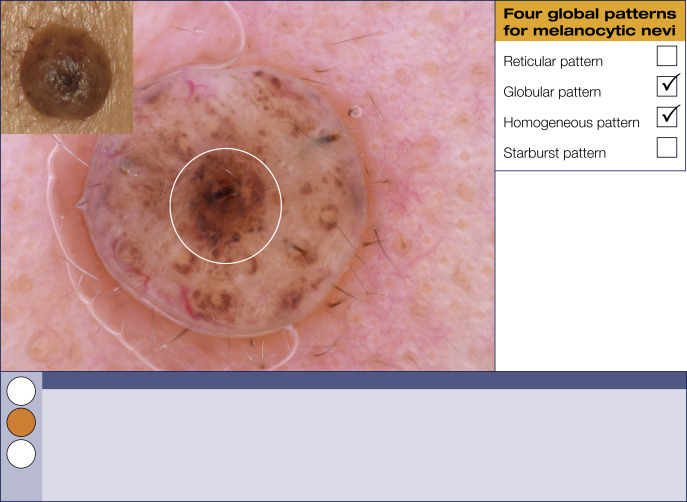
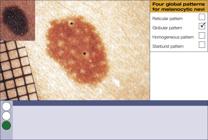
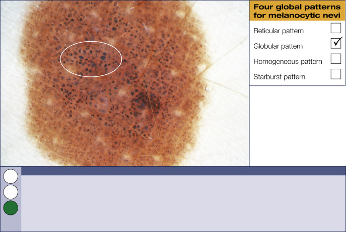
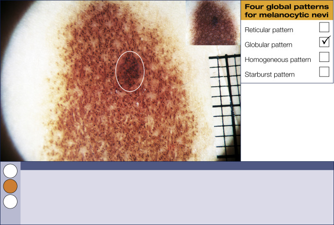
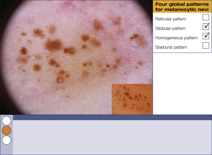
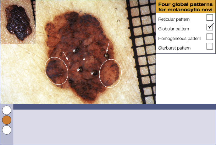
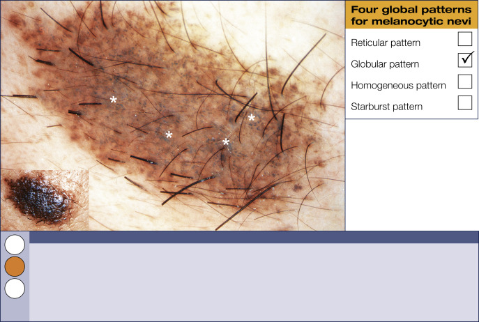
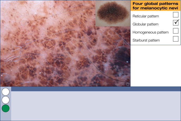
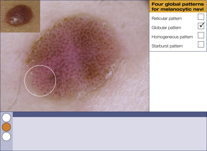
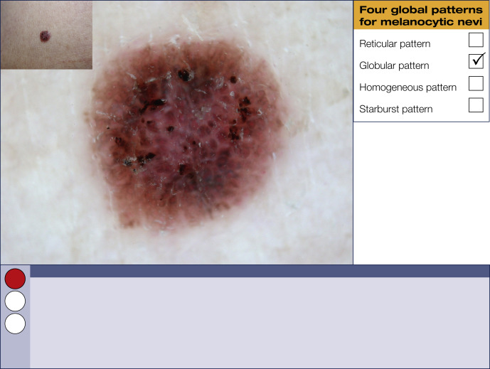

Globular pattern


Variously sized, round to oval brown structures fill these melanocytic lesions. This pattern can be found in congenital and acquired melanocytic and Clark (dysplastic) nevi.
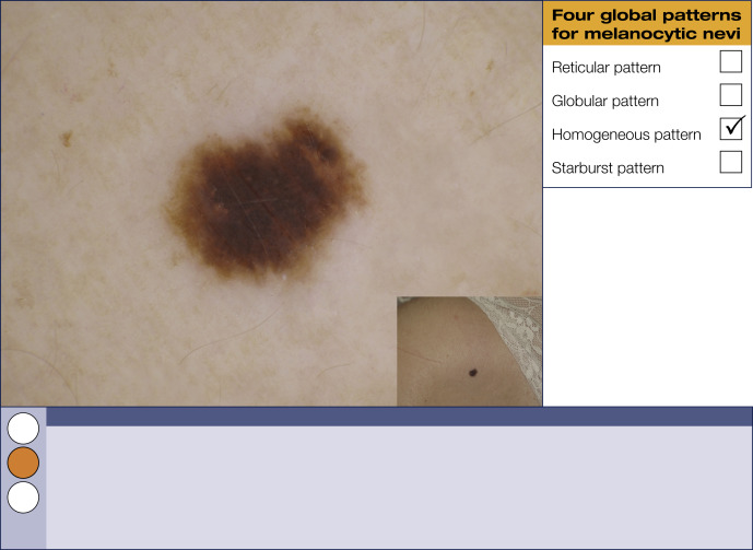
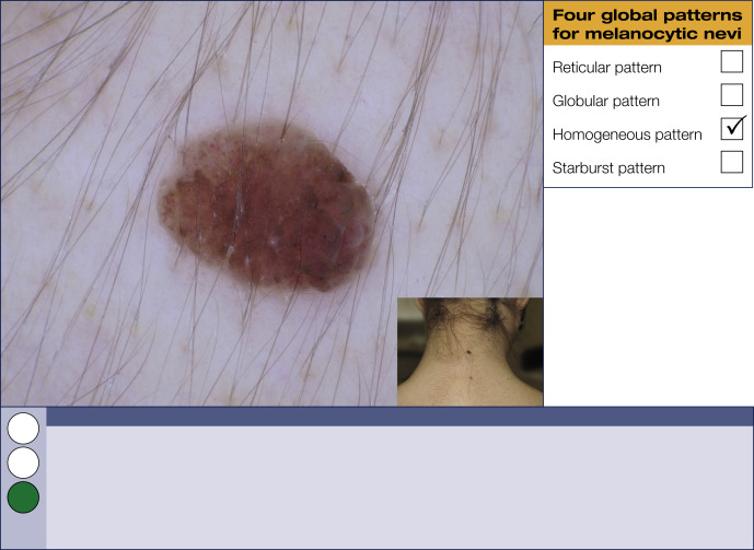
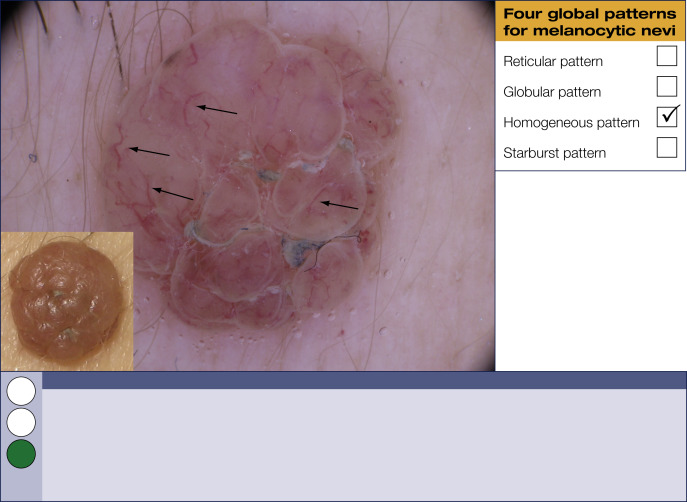
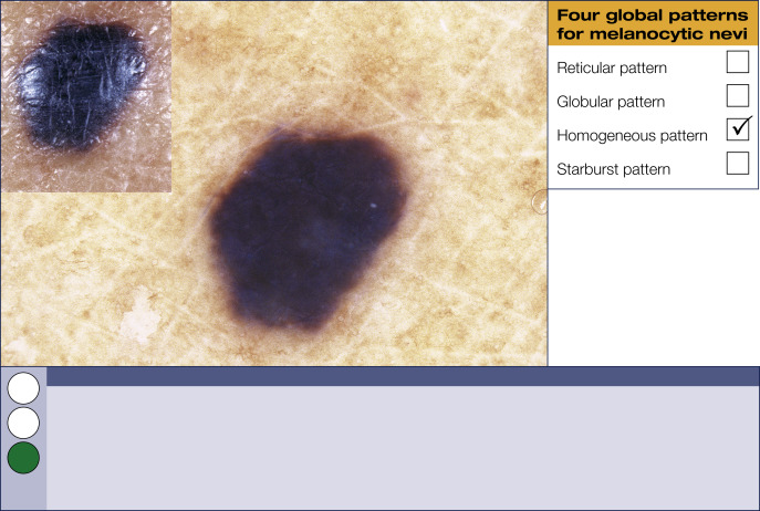
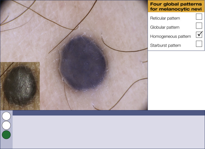
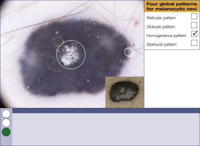
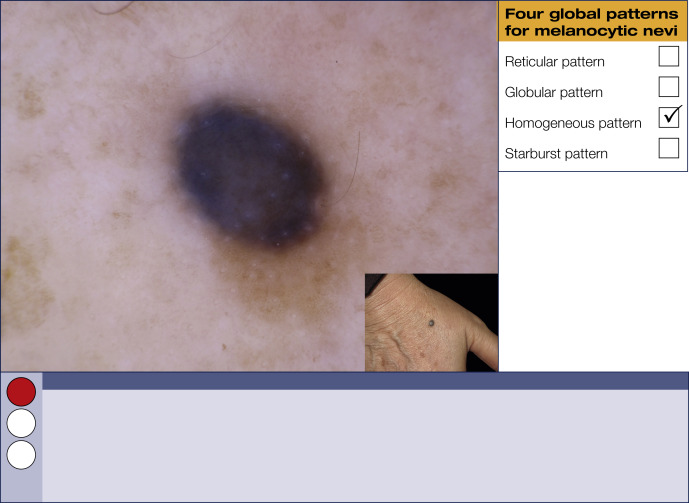
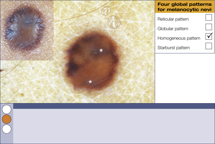
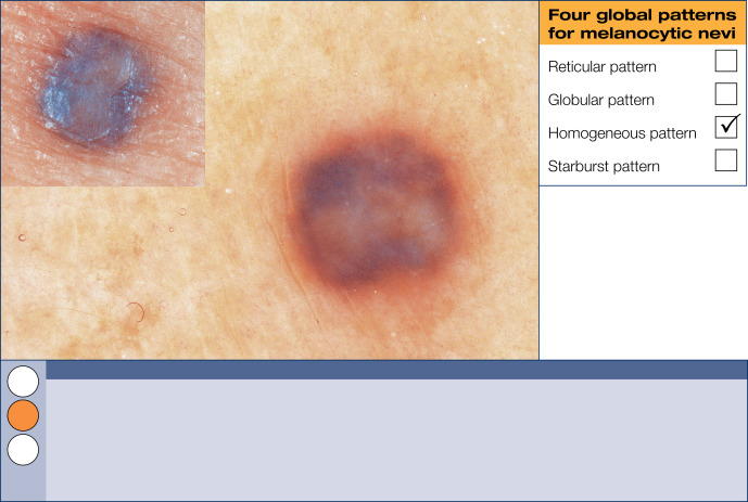
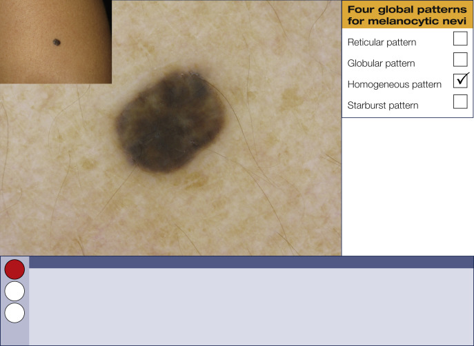
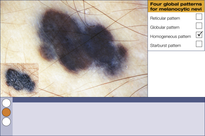

Homogeneous pattern


This pattern is characterized by a diffuse, uniform, structureless color filling most of the lesion. Colors include black, brown, gray, blue, white, or red. A predominantly bluish color is the morphologic hallmark of blue nevi.
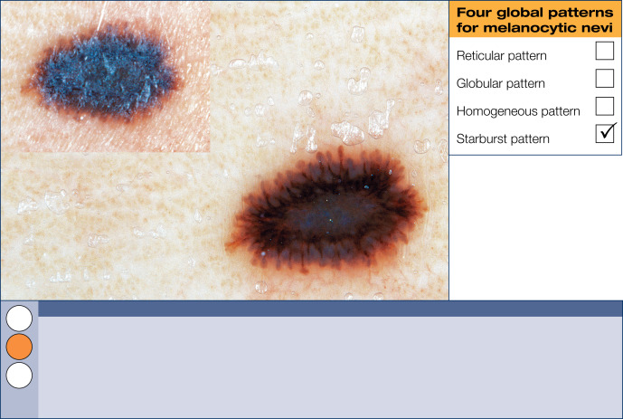
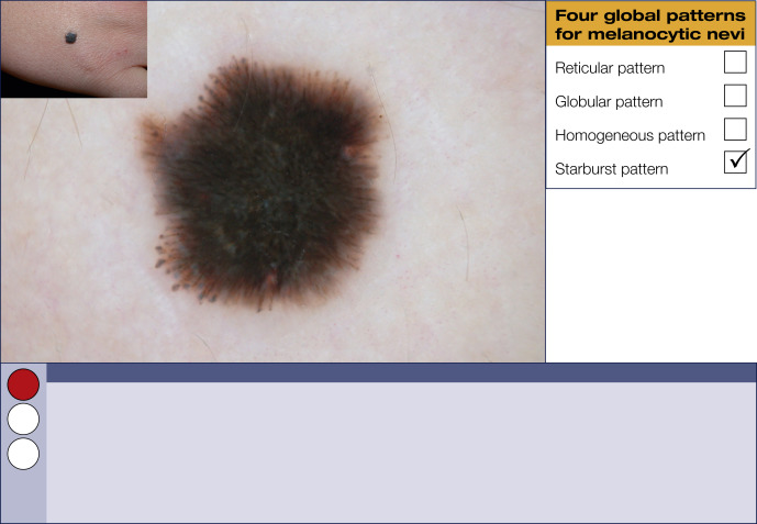

Starburst pattern


The starburst pattern is characterized by the presence of pigmented streaks and/or dots and globules in a radial arrangement at the periphery of a melanocytic lesion. This pattern is the stereotypical morphology in Spitz nevi.


Fig. 61


Nevus.

The reticular type is probably the most common dermoscopic feature of a flat acquired melanocytic nevus. It is characterized by a typical pigment network that fades out at the periphery. There are also a few small islands of hypopigmentation—a common finding in benign nevi. The histopathologic distinction between a junctional nevus and a compound nevus is commonly given, but the distinction cannot always be made dermoscopically. Moreover, it is clinically irrelevant.


Fig. 62


Nevus.

Here is another example of the morphology seen with the reticular type of banal nevus. The quality of the typical pigment network demonstrates darker and thicker lines. The benign nature of this lesion is emphasized by the fading out at the periphery of the pigment network.


Fig. 63


Nevus.

This is a reticular-type lesion with a few dots. Please note in the center of the lesion, the pigment network is lacking. In addition, there are a few brown dots (also called clods) at the periphery; an indication that this nevus is still growing. This lesion can also be called a Clark, dysplastic, or atypical nevus; it is not a melanoma.


Fig. 64


Nevus.

This lesion is characterized by a rather typical pigment network. Closer scrutiny reveals several brownish to bluish dots (clods) in the central and paracentral part of this lesion. As a general rule in reticular lesions, which are not palpable, clinically the differentiation between a junctional and a compound nevus is not possible dermoscopically.


Fig. 65


Nevus.

A reticular-homogeneous pattern, as seen here, can be seen in banal nevi. In the center, there is homogeneous black pigmentation (black lamella), and at the periphery there is an annular distribution of a typical pigment network. Once again, the pigment network fades at the periphery—a sign of a benign nature. If this was a solitary lesion, in situ melanoma would be the differential diagnosis. Most people with this dermoscopic appearance have multiple similar-appearing nevi, favoring low-risk pathology. Tape stripping can peel away the black lamella and allows one to see whether there are any underlying typical or atypical structures.


Fig. 66


Nevus.

The unusual type of reticular-homogeneous pattern seen here is more often found in younger pediatric patients. In the center of the lesion, there is homogeneous hypopigmentation (not to be confused with the bony-milky white color of regression), and this is surrounded by a small rim of pigment network. The lines of the pigment network are thickened and the meshes are slightly irregular. The overall architecture of the network, however, is symmetrical and regular.


Fig. 67


Nevus.

A stereotypical reticular pattern is seen here. The pigment network is typical, but unevenly distributed and fades out at the periphery. In addition, there are hypopigmented areas throughout the lesion ( arrows ). This nevus does not reveal criteria used to diagnose melanoma (melanoma-specific criteria). Because of the uneven distribution of the pigment network and variations in the shades of brown, the novice dermoscopist should consider excision or close dermoscopic and clinical follow-up.


Fig. 68


Nevus.

The patchy reticular pattern shown here is associated with an uneven distribution of a typical pigment network. The intensity of pigmentation of the lines alternates, giving this pigment network a patchy appearance. The general principle to remember is that any unevenness of a relatively regular-appearing pigment network is a minor cause for concern. Please note the whitish halo around a hair at the lower left pole of the lesion. This is a rather common finding in reticular nevi.


Fig. 69


Nevus.

This nevus shows a variation of reticular-pattern morphology. Note the zone of homogeneous hypopigmentation in upper half of the lesion. This is not an area of regression that would be seen in melanoma. Still, this lesion is clearly asymmetric in shape and dermoscopic structure and therefore this lesion was excised and called a high-risk nevus. Always remember: when in doubt cut it out.


Fig. 70


Nevus.

This dermoscopic picture is very worrying. The reticular pattern with eccentric hyperpigmentation dermoscopically simulates in situ melanoma arising in a pre-existing nevus. The upper right half of this lesion is characterized by a slightly atypical pigment network ( arrows ). On the left lower side, there is an area of homogeneous hypopigmentation with a few foci of delicate pigmentation commonly seen in benign nevi. Do not hesitate to excise a lesion that looks like this as soon as possible. The final histopathologic diagnosis is in situ melanoma within a pre-existing nevus in 10% of similar-appearing lesions. In this case, the diagnosis was Clark (dysplastic) nevus, compound type.


Fig. 71


Nevus.

This is a rather unusual combined nevus, with a dome-shaped globular nevus on the lower left site and a variation on the theme of a flat reticular nevus on the upper right site. This lesion should undoubtedly be excised because the differential diagnosis represents a hypomelanotic nodular melanoma arising within a superficial melanoma or a pre-existing dysplastic (Clark) nevus. However, this lesion turned out to be a dysplastic (Clark) nevus adjacent to a benign dermal nevus.


Fig. 72


Nevus.

This is most probably a nevus with a complex overall dermoscopic pattern revealing the characteristics of a reticular and a globular nevus (left pole). This lesion displays several unusual dermoscopic aspects with prominent blue-white structures (more bluish-black) in the center. Always excise a lesion like this one. The histopathology diagnosis here was a dysplastic (Clark) nevus, compound type.


Fig. 73


Nevus.

This light-brown pinkish lesion reveals a central hypopigmented homogeneous area surrounded by a subtle, not very pronounced pigment network in a ring-like fashion. The unusual aspect of this lesion is its pinkish color, and in the absence of any history of growth, annual follow-up is the management approach we choose for this patient.


Fig. 74


Nevus.

This lesion can be regarded as a typical example of a reticular melanocytic proliferation. There is a central zone of hyperpigmentation. The pigment network has rather regular qualities throughout the lesion. However, it does not thin out nicely along the periphery as commonly observed in reticular nevi. Because of this dermoscopic finding and heavy pigmentation, this is potentially a high-risk lesion. Histopathologically, this was diagnosed as a junctional type of dysplastic (Clark) nevus. Novice dermoscopists should not hesitate to excise lesions that look like this.


Fig. 75


Nevus.

This is another example of a reticular-homogeneous nevus with an annular reticular pattern in the periphery and large central homogeneous hypopigmented area. The color of the hypopigmented area is not bony-white as observed in regressive melanoma, and because of the overall symmetry of this lesion, annual follow-up can be advised confidently by the novice dermoscopist.


Fig. 76


Nevus.

This lesion has a uniform reticular pattern with only a delicate focus of paracentral hyperpigmentation ( circle ). The pigment network is typical and slightly fades out at the periphery. The overall shape of the lesion, however, is a bit asymmetric and lesions like this one should always be followed up.


Fig. 77


Nevus.

This is another example of the protean variation of morphology within melanocytic proliferations exhibiting the reticular pattern. In contrast to Fig. 76 , the pigment network here is mostly atypical with a tendency to stop abruptly at the periphery. This high-risk nevus cannot be distinguished dermoscopically from a superficial melanoma (or an in situ melanoma) and needs to be excised.


Fig. 78


Nevus.

This is a predominantly reticular type of nevus with a pigment network fading out nicely at the periphery. In our opinion, this is the most common type of benign nevus in men. Still, as a rule, we do recommend annual follow-up and self-monitoring of reticular nevi.


Fig. 79


Nevus.

This shows a stereotypical globular pattern of a benign nevus. There are numerous dots and globules of similar shape and varying size throughout the lesion. No melanoma-specific dermoscopic criteria are seen. This pattern is most commonly seen in adolescents but can also be found in adults. The histopathology could show a junctional or compound nevus.


Fig. 80


Nevus.

This shows one of the many variations of the morphology seen with the globular pattern. The most relevant aspect of this lesion is the even distribution of closely packed, similar-appearing dots and globules. In addition, there are a few milia-like cysts in the center of the lesion ( arrows ). Milia-like cysts are not seen only in seborrheic keratosis.


Fig. 81


Nevus.

This globular pattern shows dots and globules that are not closely packed together, are similar in size and shape, and have a slightly uneven distribution. Please note an increase of globules at the periphery of the lesion (peripheral rim of globules). This specific pattern is commonly observed in symmetrically growing nevi. No melanoma-specific criteria are seen in this otherwise banal lesion.


Fig. 82


Nevus.

Most of this lesion is characterized by a homogeneous brownish pigmentation and numerous subtle dots and globules. The slightly increased focal pigmentation at 2 to 3 o’clock is a common finding in benign nevi. Please remember nothing in nature is completely symmetrical.


Fig. 83


Nevus.

This image shows a more worrisome variation of the globular pattern. Numerous dots and globules are unevenly distributed throughout the lesion ( circle ) and vary in size and shape.


Fig. 84


Nevus.

Here is another globular type of nevus. Numerous light-brown to blue-gray dots and globules, which are of similar size and shape, are distributed regularly throughout the lesion. The only worrisome area is a collection of about 15–20 gray globules ( circle ), which prompted the excision of this compound type of Clark (dysplastic) nevus. Study lesions carefully to look for subtle yet potentially high-risk criteria.


Fig. 85


Nevus.

This is another stereotypical example of the globular pattern of nevus, in which the globules are very easy to see. Throughout this lesion several dark-brown dots and globules display a rectangular shape (cobblestone-like). This morphologic phenomenon is often found in congenital or congenital-like nevi. Dermoscopically, this lesion gives the impression of a papillomatous or raised character. Histopathologic examination revealed a predominantly dermal compound nevus.


Fig. 86


Nevus.

The globular pattern seen here is very similar to that in Fig. 85 . The lesion is composed of closely packed dots and globules characterized by various shades of brown. In addition, there are several whitish dots and globules representing keratin accumulation. The variation of the color might alarm the inexperienced dermoscopist. Remember, if in doubt, cut it out. This was a benign, mostly dermal nevus. After seeing and excising a few lesions with this dermoscopic appearance, the dermoscopist will feel more comfortable about not excising lesions that look like this.


Fig. 87


Nevus.

This lesion shows again a globular pattern. It contains numerous brown to gray globules, which are evenly distributed throughout the lesion. The gray globules are situated predominantly in the center of the lesion and correspond to nests of pigmented nevus cells in the papillary dermis. Remarkably, globular nevi represent the stereotypical nevus subtype among children and teenagers.


Fig. 88


Nevus.

It is amazing to see the many different variations on the theme of globular nevi. In the previous pages, we have seen quite a few benign globular nevi, but all are morphologically different and unique. The striking aspect of this uniformly pigmented globular nevus is its dark brown pigmentation. We are happy to follow this nevus and recommend self-monitoring.


Fig. 89


Nevus.

This globular nevus raises at least the orange flag because the globules composing this lesion vary slightly in size, shape, and color and are also slightly unevenly distributed throughout the lesion. In addition, this lesion has an incomplete rim of peripheral globules, indicating that this lesion will continue to grow. Because the patient was concerned about this lesion, a deep shave biopsy was performed. The final histopathologic diagnosis was a compound type of dysplastic (Clark) nevus.


Fig. 90


Nevus.

Numerous irregularly sized brownish dots and globules are seen throughout this lesion. Although it is rather small, the dermoscopic asymmetry is striking. The pinkish color is an important clue that this could be a high-risk lesion. Because of its high-risk appearance, a lesion like this one warrants a second histopathologic opinion if it is signed out as a benign nevus as was the case here.


Fig. 91


Nevus.

One has to look carefully to recognize that this heavily pigmented nevus reveals a globular and not a homogeneous pattern in its central part. The lighter pigmented peripheral ring displays a pattern reminiscent of globules and reticulated lines telling us that in morphology there is always an overlap of features. We were confident that this lesion was a variation on the theme of a benign globular nevus and recommended annual follow-up and self-monitoring.


Fig. 92


Nevus.

In some instances, the distinction between a globular and a reticular nevus is not that easy, as evidenced by this slightly irregularly outlined lesion. In several parts, a pigment network characterized by thickened lines and small holes prevail, whereas, toward the periphery, a more globular pattern becomes evident. Although we are raising the orange flag here, the final management decision needs to be done in context with the clinical setting. We have excised this lesion and the histopathology has ruled out a melanoma.


Fig. 93


Nevus.

This lesion is characterized by diffuse homogeneous pigmentation. There is a subtle rim of radially oriented line segments at the periphery at 9 o’clock and subtle blue-white structures in the center. The dermoscopic differential diagnosis includes Clark (dysplastic) nevus and Spitz nevus. We raised the orange flag and excised this lesion. The lesion was reported as a compound type of Clark (dysplastic) nevus with so-called spitzoid features.


Fig. 94


Nevus.

Apart from the blue-white structures and tiny dots in the central part ( circle ), this lesion displays a rather uniform subtle reticular pattern, which made us comfortable to follow up this lesion. We are well aware that some colleagues would prefer to excise a lesion like this one for peace of mind. Also the clinical image was reassuring for us that we were dealing with a nevus.


Fig. 95


Nevus.

This lesion is characterized by a reticular-homogeneous pattern. Please note the focus of atypical pigment network ( circle ). In addition, the left lower part of the lesion exhibits blue-white structures, and these two signs are sufficient to warrant excision. In the realm of dysplastic (Clark) nevus, it is difficult to determine whether a lesion is low or high-risk dermoscopically; therefore, the novice is best advised to excise gray-zone lesions as this one.


Fig. 96


Nevus.

This is another example of a benign globular nevus with globules slightly varying in size, shape, and coloration. Despite the irregular outline of this lesion, no action but follow-up has to be undertaken.


Fig. 97


Nevus.

This is a rather commonly observed variation on the theme of a reticular type of nevus. These lesions are frequently found in adults. We judge this pigment network as somewhat atypical and despite rather uniformly distributed in this roundish lesion, we raise the orange flag. It is also not really fading out at the periphery, which is rather infrequently seen in reticular nevi. Without any specific history of growth by the patient, we were happy with short-term follow-up of this nevus and, in addition, recommended self-monitoring.


Fig. 98


Nevus.

This lesion is a variation of the homogeneous-reticular type of nevus reminiscent of a so-called black nevus. Multiple jet-black homogeneous zones are seen at the periphery. Use tape stripping for this black lesion mimicking in situ melanoma.


Fig. 99


Nevus.

This is a dome-shaped melanocytic nevus that reveals a subtle globular pattern with numerous light-brown dots and globules throughout. Multiple blood vessels with dotted ( asterisks ) and comma-like appearances ( arrows ) are seen. There are also a few milia-like cysts ( circles ), but this is not a seborrheic keratosis. Clinically this lesion could be confused with a basal cell carcinoma, but the vessels in a basal cell carcinoma are thick and branched (arborizing) and there would be no yellow color.


Fig. 100


Nevus.

This lesion has a globular pattern containing numerous brownish-blue dots and globules, which vary in size and shape, and a central irregular brownish blotch ( circle ).


Fig. 101


Nevus.

This clinically broad sessile nodule has a papillomatous surface and a few irregularly shaped comedo-like openings ( arrows ). Sometimes it is not possible to differentiate the comedo-like openings from globules. The thin pigmented lines are not pigment network but pigmentation in the furrows of the lesion. The soft, compressible nature points to it being low risk. Palpate suspicious lesions, but if in doubt, cut them out.


Fig. 102


Nevus.

This is another broad, sessile nodule characterized by a papillomatous surface. There are some comedo-like openings ( arrows ) and a few bluish dots and globules ( asterisks ). These can be confused with blue-white structures.


Fig. 103


Nevus.

Here is another papillomatous dermal nevus. Please note the subtle variation of gray and brown colors throughout this slightly papillomatous nodule. Also, the presence of numerous terminal hairs within this nevus is rather typical for a benign lesion. This is another lesion to palpate. Compressibility and easy movement from side to side are good clinical signs in favor of its benign nature.


Fig. 104


Nevus.

It is common to see a papillomatous nevus ( circle ) in transition with a flat melanocytic nevus ( solid arrow ). The flat component can give a worrisome clinical appearance, which in most cases is not high risk when viewed with dermoscopy. The dome-shaped nodule is characterized by numerous comedo-like openings ( asterisks ). In addition, there are comma-like vessels ( open arrows ) throughout the lesion. Comma-shaped vessels are not characteristically seen in melanomas. At the lower margin, there is a flat brownish area with regular dots and globules.


Fig. 105


Nevus.

This papillomatous dermal nevus is relatively featureless, but the blood vessels ( arrows ) might make one consider basal cell carcinoma in the differential diagnosis. The vessels of basal cell carcinoma, however, are linear, sharp in focus, and branched (arborizing). This elevated papillomatous nodule reveals a homogeneous pattern and has a light-brown color. Commonly these nevi are irritated due to incidental traumas.


Fig. 106


Nevus.

In this bizarre dermoscopic picture, there are several densely aggregated exophytic papillary structures and ridges, which look like globules. There are also a few irregular crypts and furrows ( arrows ), which represent a variation of the morphology seen with comedo-like openings. In the center, there is an accumulation of yellowish-white keratotic material ( asterisks ). Palpate this lesion and it will be soft, which will be one criterion in favor of it being a banal nevus.


Fig. 107


Nevus.

This lesion is similar to that in Fig. 106 and is composed of densely aggregated exophytic papillary structures intermingled with furrows ( asterisks ). In addition, there are a few regular brown dots and globules ( arrow ) and blue-white structures. A small banal reticular-type nevus is seen in the right lower corner.


Fig. 108


Nevus.

This elevated nevus on the forehead is characterized by some light- to dark-brown dots and globules, particularly in the center of the lesion ( circle ). Please note the presence of roundish holes representing hair follicles. Closer scrutiny shows hairs in the center of a few follicles. These nevi are so often inflamed due to ingrown hairs and ruptured hair follicles. Because of the relatively pronounced pigmentation, we raised the orange flag. This benign nevus was excised as requested by the patient.


Fig. 109


Nevus.

Here is a very subtle type of globular pattern in a flat melanocytic nevus with numerous tiny dots and multifocal hypopigmentation ( asterisks ). This pattern can be seen with congenital or Clark (dysplastic) nevi.


Fig. 110


Nevus.

This image shows one of the stereotypical patterns seen with congenital nevi. It is a reticular pattern with islands of light, featureless color similar to those seen in Fig. 109 , but more dramatic. The pigment network in the central portion is more heavily pigmented and the lines are thickened when compared to those at the periphery. There is also a focus of blue-white structures ( arrow ). Commonly, congenital nevi look worrisome with dermoscopy but not histologically. Islands of normal skin + islands of criteria = congenital melanocytic nevus.


Fig. 111


Nevus.

The globular pattern seen here is intermingled with roundish white holes and is characterized by numerous tiny bluish dots and globules ( circle ) situated predominantly in the center of the lesion, and many light-brownish globules peripherally. The overall dermoscopic architecture of this lesion is symmetrical and regular, and excision is not indicated.


Fig. 112


Nevus.

This lesion shows another of the many variations of the globular pattern with numerous hypopigmented roundish areas. This pattern is very suggestive of a congenital melanocytic nevus. Numerous brownish dots and globules are evenly distributed throughout the lesion. In the upper section, there is an oval dark-brown pigmented area. This blotch ( circle ) could represent high-risk pathology, and for this reason, the lesion should be excised.


Fig. 113


Nevus.

This lesion with a particular homogeneous pattern is a congenital speckled nevus, also called nevus spilus. There is a rather characteristic pattern of several brownish homogeneous dots and clods on a homogeneous light brown to skin-colored background. The novice may be confounded by this lesion and consider an unusual melanoma in the differential diagnosis. We felt confident to recommend follow-up and self-monitoring of this special type of congenital nevus.


Fig. 114


Nevus.

This papillomatous nevus is composed of a few exophytic papillary structures ( circles ) and some comedo-like openings ( asterisks ). In addition, there are a few milia-like cysts ( arrows ) and blue-white structures. If a worrisome-looking lesion like this is palpated, it should be soft and compressible—this sign indicates that it is benign.


Fig. 115


Nevus.

This reticular nevus is characterized by a pigment network pattern that immediately raises our suspicion. There are certainly areas with a thickened and branched pigment network ( circles ) and also the other parts of the pigment network reveal some features of irregularity. This dysplastic (Clark) nevus simulates in situ melanoma and should be excised.


Fig. 116


Nevus.

This nevus is characterized by the presence of numerous hairs, which is diagnostic of a congenital melanocytic nevus. There are also brownish globules throughout the lesion intermingled with numerous small blue dots ( asterisks ), which represent collections of melanophages in the papillary dermis and raise the suspicion of a regressing melanoma. Against the dermoscopic diagnosis of melanoma are the presence of multiple hairs and symmetry of color and structure.


Fig. 117


Nevus.

Here is another example of a nevus with dark hairs, which might be best interpreted as a small congenital melanocytic nevus on the face. The dermoscopic hallmark of this lesion is a regular pseudopigment network formed by numerous round areas, which represent follicular openings. This criterion is site specific. Because of the dermoscopic symmetry of color and structure, a melanoma can be ruled out with certainty. Pigment network is not the same as pseudopigment network.


Fig. 118


Nevus.

This is a stereotypical example of a nevus spilus, which is characterized by several foci ( circles ) of a subtle brownish pigment network on a diffuse light-brown background. Each of these spots is reminiscent of a reticular type of nevus. Melanoma can develop in a nevus spilus; therefore, dermoscopy is a useful tool for examining these lesions. Look for the same high-risk criteria as for other types of melanocytic nevi.


Fig. 119


Nevus.

This nevus exhibits a reticular-homogeneous pattern, with the homogenous zone in the center of the lesion. The periphery is characterized by a regular patchy distribution of small foci of typical pigment network arranged in an annular pattern. This lesion reveals a very distinct “Gestalt” called a cockade and is nowadays commonly termed targetoid or cockade nevus (nevus en cocarde). Because of the overall symmetrical aspect of this lesion, we are raising the green flag. Of course, monitoring of this lesion and annual follow-up is recommended.


Fig. 120


Nevus.

At higher magnification, this congenital nevus has a very worrisome appearance dermoscopically because there is asymmetry of color and also of structure. However, in melanocytic lesions larger than 1 cm in diameter, we have always taken into consideration the clinical appearance. It is well known that dermoscopy of congenital nevi may be confounding and lead us astray. Putting together the clinical and the dermoscopic features of this lesion, we are confident to follow up this congenital nevus.


Fig. 121


Nevus.

This is a stereotypical example of a blue nevus characterized by diffuse homogeneous pigmentation. There is also a small rim of brownish pigmentation. The differential diagnosis of this blue nevus is a hemangioma and nodular or cutaneous metastatic melanoma. The history of the lesion is vital to make the correct dermoscopic diagnosis.


Fig. 122


Nevus.

This Fig. shows a stereotypical example of a blue nevus. Note the homogeneous blue pigmentation throughout the lesion, a morphologic finding that is observed basically only in blue nevi and in tattoos. Very rarely you can find a similar dermoscopic appearance in rapid-growing cutaneous melanoma metastases. In the latter instance, patient history data will lead you to the diagnosis of melanoma metastasis.


Fig. 123


Nevus.

This image shows another example of a typical blue nevus. The whitish area in the center of the lesion ( circle ) is just a scale. If there is no history of growth, we can confidently raise the green flag here.


Fig. 124


Nevus.

This is a blue nevus with a central whitish zone simulating a regressive Clark (dysplastic) nevus or even a regressing melanoma. There is also an off-center light brown homogenous zone. The brown zone, set asymmetrically to the main body of the lesion, may concern dermoscopists. Remember, if in doubt, cut it out, and this is particularly relevant for nodular lesions. We decided to completely excise this lesion, and the histopathologic diagnosis here was a cellular blue nevus.


Fig. 125


Nevus.

This blue nevus is a predominantly firm nodule with a smooth surface. The clinical differential diagnosis includes hypomelanotic melanoma, dermatofibroma, or dermal nevus. The nevus has a diffuse light-brownish color bordered by small zones of darker pigmentation and blue-white structures ( asterisks ). No other dermoscopic criteria are seen. Because a hypomelanotic melanoma cannot be ruled out with certainty, a lesion with this dermoscopic picture should be excised.


Fig. 126


Nevus.

This variation of the morphology seen in blue nevi simulates hypomelanotic melanoma and is characterized by a fusion of diffuse bluish and whitish zones. There is a complete lack of individual dermoscopic criteria.


Fig. 127


Nevus.

This is an example of an unusual blue nevus with asymmetric zones of bluish-brown pigmentation and whitish zones. A lesion like this one raises important differential diagnostic considerations, such as Spitz nevus and nodular melanoma. Although there is a small probability that this is indeed a nodular melanoma, we are raising the red flag here.


Fig. 128


Nevus.

This lesion is a variation of a blue nevus. Dermoscopically, it is characterized by homogeneous blue and gray color surrounded by a faint ring of lighter blue color. There are no hints of local dermoscopic features, particularly melanoma-specific criteria. Nevertheless, because of the lesion’s asymmetry of contour and color, excision is justified to rule out a melanoma. The history is also an important factor in this case.


Fig. 129


Nevus.

This is a stereotypical example of a Spitz nevus with a starburst pattern. There is a symmetrical ring of streaks around the entire lesion and a central blue-white structure. Both these dermoscopic features are commonly found in Spitz nevi. If the streaks are not at all areas of the periphery, it could be the dermoscopic picture of a melanoma. A starburst pattern should immediately make one think of Spitz nevus.


Fig. 130


Nevus.

This lesion is also reminiscent of a Spitz/Reed nevus and is similar to that in Fig. 129 , but with asymmetrically distributed pseudopods at the periphery. It also has the starburst pattern with a central blue-white structure. As a rule, excision of a lesion with this dermoscopic appearance is recommended, particularly if the individual is over 14 years of age.


Fig. 131


Nevus.

This is predominantly a globular type of Spitz nevus, which dermoscopically raises a suspicion of melanoma. It is a relatively symmetrical lesion characterized by numerous brown to bluish globules rather evenly distributed throughout the lesion. In the left lower corner of the lesion, there are several dotted vessels ( circle ). The decision about whether to closely follow or excise a lesion that looks like this depends on the clinical setting.


Fig. 132


Nevus.

This lesion is similar to the one before, but the dots and globules are more prominent. This globular pattern can be seen in banal, dysplastic (Clark), and Spitz nevi as well as rarely in melanomas. In addition, there are hints of streaks in the periphery (1–2 o’clock) and a so-called negative pigment network in the central parts of the lesion, both features suggestive of a Spitz nevus. There are also several black dots throughout the lesion, making this lesion quite suspicious for melanoma. Because of the equivocal dermoscopic appearance, this lesion was excised and diagnosed histopathologically as a Spitz nevus.

Only gold members can continue reading. Log In or Register to continue

Stay updated, free articles. Join our Telegram channel

Mar 31, 2020 | Posted by in Dermatology | Comments Off on Pattern analysis: Dermoscopic criteria for specific diagnoses

Full access? Get Clinical Tree

Get Clinical Tree app for offline access