Partial Mastectomy
Sheryl G. A. Gabram
 Indications/Contraindications
Definitions
Breast cancer surgery is trending in the direction of “less is more” as the number of patients newly diagnosed qualify for breast conservation surgery over mastectomy and sentinel lymph node biopsy instead of complete axillary lymph node dissection. Breast conservation treatment refers to removal of the breast cancer combined with radiation therapy to offer optimal local control, usually performed in association with axillary surgery. The surgical removal of the cancer from the breast may be referred to as lumpectomy, partial mastectomy, or quadrantectomy. Lumpectomy and partial mastectomy are essentially synonymous terms and indicate surgical removal of the tumor with the intent of obtaining a clear margin. In the United States, there is no ICD-9 code (Internal Classification of Diseases, 9th revision codes used for billing purposes) for lumpectomy, and therefore when reporting the operative details of performing breast sparing surgery, many surgeons will use the term “partial mastectomy” with ICD-9 code 19301. Quadrantectomy involves removal of a larger portion of breast tissue, a quadrant, often with overlying skin and removal of the breast tissue posteriorly from the pectoralis muscle. The latter technique has been popularized in Europe while in the United States, partial mastectomy/lumpectomy is performed more commonly.
Safety
The use of breast conservation treatment for early stage breast cancer has been tested in 6 worldwide prospective randomized clinical trials comparing lumpectomy/partial mastectomy (or quadrantectomy) to mastectomy. The long-term data indicate that when a negative margin is obtained, survival of breast sparing surgery compared to mastectomy is identical. In the United States National Surgical Adjuvant Breast and Bowel Project (NSABP) B-06 clinical trial, while the overall long-term survival was similar for those patients treated with lumpectomy, mastectomy, and lumpectomy with radiation, local control was markedly improved for those patients treated with radiation (in breast recurrence for lumpectomy with radiation of 14.3% vs. 39.2% for lumpectomy without radiation). Recurrence after mastectomy in this large trial was 10.2%, not significantly different for those patients treated with lumpectomy and radiation. The randomized trial from Milan on quadrantectomies also supports the safety of breast conservation for those patients meeting criteria for this procedure.
Indications
The indications for breast-sparing surgery include the following:
Patients willing to receive radiation therapy with no contraindications
Reasonable breast to tumor size ratio that would yield an acceptable cosmetic result after breast sparing surgery
Localized disease assessed via breast imaging and clinical examination (disease limited to one quadrant)
Contraindications
The contraindications for breast sparing surgery include the following:
Patient not a candidate for radiation therapy (such as those patients with scleroderma or other connective tissue diseases associated with skin involvement)
Patient with history of prior therapeutic irradiation to the breast
Patient unwilling to undergo radiation therapy
Patient requiring radiation during pregnancy (pregnancy is an absolute contraindication to receiving radiation during pregnancy, thus creating a relative contraindication to Breast Conservation Therapy (BCT) during first and second trimesters)
Two or more tumors in separate quadrants of the breast
Evidence of diffuse breast involvement by imaging or clinical examination
Persistently positive margins despite attempts at reasonable surgical re-excision
Large tumor that would preclude a reasonable cosmetic outcome
Imaging
The clinical examination and imaging appearance of the tumor will determine whether breast conservation is feasible. It is important for surgeons to review all imaging to confirm localized disease that may or may not be associated with clinical findings. If the tumor is not palpable, a preoperative wire localization is necessary to identify the epicenter of the tumor for excision. Localization may also be necessary if the tumor is vaguely palpable, again to minimize the amount of breast tissue removed.
Increasingly, breast magnetic resonance imaging (MRI) is performed to determine the extent of the disease process especially in younger women as well as those women with extremely dense or heterogeneously dense breast tissue, where extent of the disease process may be underestimated by routine mammography. Some studies indicate that the use of this modality will change the operative plan in 16% of cases, with additional tumor identified in a multicentric fashion or the primary larger than suspected on routine mammography. The downsides of using breast MRI routinely is that no data have shown an improvement in survival for those patients who clinically are candidates for breast conservation. In the United States, insurance companies may not cover the charge of an MRI for all newly diagnosed breast cancer patients. Because the specificity is low, often a patient’s surgery will be delayed if abnormalities on a breast MRI are uncovered that necessitate biopsy to either prove or disprove multicentricity. Currently, not all surgeons are advocates of routine MRI in planning for partial mastectomy.
Neoadjuvant Therapy (Systemic or Hormonal)
Another trend in breast cancer treatment is targeted individualized therapy depending on various tumor characteristics. This had led to an increase in delivering preoperative systemic chemotherapy or hormonal therapy to better document the treatment effect of a specific regimen prior to surgery. Performing surgery after neoadjuvant therapy will contribute significant information on tumor responsiveness of a particular regimen and
help guide future systemic treatment. In addition, some patients will become candidates for breast conservation if treated with systemic therapy prior to surgery. Studies have shown that it is safe to offer upfront therapy (neoadjuvant chemo or hormonal treatment, also referred to as primary systemic chemotherapy or hormonal therapy) with only a minority of tumors progressing on therapy during such an approach. It is important for surgeons to clinically follow patients during neoadjuvant therapy to assess response to treatment and ensure that unresponsive tumors do not require a change in therapeutic intervention that may include a more immediate surgical procedure. Preoperative hormonal therapy is used for elderly patients and may take the place of surgery in a patient with multiple comorbid diseases who may be at high risk for surgery. For this elderly group, each case is decided upon on an individual basis and if the patient responds well to this approach, consideration for partial mastectomy in an outpatient setting under intravenous sedation in the operating room can take place at the point the tumor is no longer responding to the treatment regimen.
help guide future systemic treatment. In addition, some patients will become candidates for breast conservation if treated with systemic therapy prior to surgery. Studies have shown that it is safe to offer upfront therapy (neoadjuvant chemo or hormonal treatment, also referred to as primary systemic chemotherapy or hormonal therapy) with only a minority of tumors progressing on therapy during such an approach. It is important for surgeons to clinically follow patients during neoadjuvant therapy to assess response to treatment and ensure that unresponsive tumors do not require a change in therapeutic intervention that may include a more immediate surgical procedure. Preoperative hormonal therapy is used for elderly patients and may take the place of surgery in a patient with multiple comorbid diseases who may be at high risk for surgery. For this elderly group, each case is decided upon on an individual basis and if the patient responds well to this approach, consideration for partial mastectomy in an outpatient setting under intravenous sedation in the operating room can take place at the point the tumor is no longer responding to the treatment regimen.
Role of Oncoplastic Surgical Techniques
Depending on the size of the partial mastectomy resection, some patients may benefit from oncoplastic surgical techniques in repairing the defect. This is an emerging field that offers patients an alternative to mastectomy, especially for very large defects in patients with generous breast tissue who may benefit from opposite breast reduction surgery (Fig. 14.1).
Factors involved in the surgical decision making for oncoplastic procedures after the performance of partial mastectomy include the following:
Timing of reconstruction in relation to radiation therapy: It is ideal to consider oncoplastic surgery prior to partial mastectomy and radiation. Patients who present after completion of surgery and radiation are a challenge because if the defect is large, they may require transfer of significant amount of autologous tissue. In this setting, the remaining breast tissue has been exposed to radiation and wound healing may be
compromised. Often these patients have chosen breast conservation therapy because of a desire to have less versus more surgery. Sometimes, completion mastectomy and immediate reconstruction are the best options for significant defects.
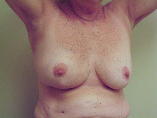
Figure 14.1 Right breast cancer with bilateral reduction surgery.
Status of tumor margins: Oncoplastic techniques can be performed immediately after the partial mastectomy or delayed a week or two for final margin status determination (but before radiation is administered). If multicentric disease is suspected, the latter approach, with delay to confirm final margins, is advantageous. However, if disease appears localized, a unified procedure can be recommended, thus avoiding a second procedure in the operating room.
Extent of breast skin and tissue resection: This will be determined by the size of the tumor and proximity to the anterior skin margin. If reduction procedures are planned, the incisions for the partial mastectomy portion of the procedure can be in the same location of the reduction incisions. Careful planning between the surgical oncologist and the plastic surgery team is necessary for this approach. At the time of surgery, specimen mammography may be necessary to confirm that the entire lesion is resected with a visually acceptable margin.
Breast size: Patients with pendulous breasts may benefit from oncoplastic techniques even if the lesion for resection is small. Radiation to large breasts can be compromised and uneven dosages may occur in this group of patients. It is desirable to perform the partial mastectomy and bilateral reduction prior to radiation therapy as opposed to performing reduction surgery after the patient has completed all therapy.
Ultimate cosmetic outcome: An assessment needs to be made preoperatively if the patient would benefit from completion mastectomy versus wide partial mastectomy and reduction of the involved breast. If a patient does not meet criteria for postmastectomy radiation, the advantage to simple mastectomy and reconstruction may be to avoid radiation. Again, a co-ordinated informed discussion with the surgical oncologist, plastic surgeon, and patient taking into account the patient’s expectations is necessary.
Patient Consideration
A detailed family history (three generations on both the maternal and paternal lineages) is necessary to determine if a patient may be at risk for the BRCA1/BRCA2 genetic mutation. Characteristics of those at increased risk for BRCA1/BRCA2 or other high penetrance mutations (p53 or PTEN) are displayed in Table 14.1. While breast conservation therapy is still an option for a patient diagnosed with the BRCA1/BRCA2 genetic mutation, the role of bilateral mastectomies should be considered as well, because these patients are at higher risk for the development of new primaries on the involved side as well as contra lateral breast cancer. Genetic testing may delay surgery; however, if a patient is a candidate for neoadjuvant therapy, that treatment can proceed while the patient is referred for genetic counseling and possible testing.
Patients will often have predetermined desires regarding breast conservation surgery versus mastectomy. Data regarding the safety of breast conservation should be discussed with the patient. Even after these discussions, a group of patients will desire mastectomy and furthermore even request bilateral mastectomies (contralateral prophylactic mastectomy) with or without immediate reconstruction. While there is no survival advantage to this approach, an increasing trend of bilateral mastectomies in the setting of unilateral cancer has been identified.
Prevention of Acute Complications
There is ongoing debate about the role of prophylactic antibiotics for breast surgery. Several studies report the advantage of prophylactic antibiotics given as a one time dose 30 minutes prior to surgery, yet others have shown no benefit. High-risk patients, those who are obese, the elderly, and diabetic may be those who benefit from prophylactic antibiotics to a greater extent. Foreign body placement, wire localizations and clips for marking biopsy sites, may add to risk of infection and one may consider use of prophylactic antibiotics in these settings as well.
Table 14.1 Indications for Referral to Genetics Counseling | ||
|---|---|---|
|
Prevention of bleeding and hematoma formation can take place by obtaining an accurate preoperative history of use of blood thinners such as daily aspirin or anti-inflammatory medications. If possible, these medications should be stopped 10 days to 2 weeks prior to surgery. Clopidogrel (Plavix) should be stopped a full week prior to surgery. Surgery should be delayed about 4 weeks after the last infusion of bevacizumab (Avastin) because of the potential of bleeding and dehiscence. Other agents that contribute to bleeding in breast patients are Vitamin E, ginseng, ginko biloba, and garlic. There is also a debate about closure of the partial mastectomy cavity. The conventional approach is double layer skin closure without deep sutures. Some surgeons prefer deep closure to decrease risk of hematoma formation, but this may compromise cosmetic appearance.
Unique Clinical Circumstances
Related posts:
Stay updated, free articles. Join our Telegram channel

Full access? Get Clinical Tree


