Parasitic Infestations, Stings, and Bites


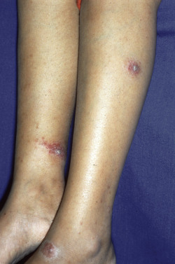
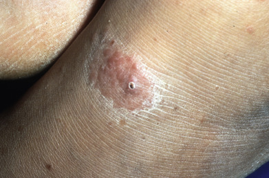
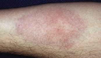
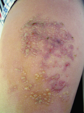
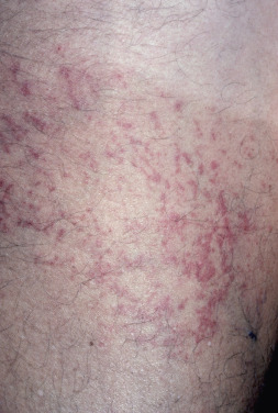
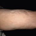
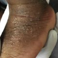
Bites and infestations present with wide-ranging manifestations, including papules, vesicles, excoriations, and urticarial lesions. Bedbug bites often involve the arms and develop clinical features of prurigo nodularis. A biopsy will demonstrate a wedge-shaped perivascular lymphoid infiltrate with endothelial swelling and eosinophils, suggesting the correct diagnosis.


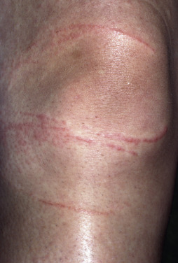
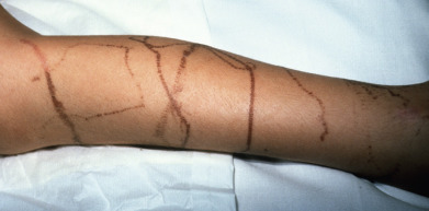
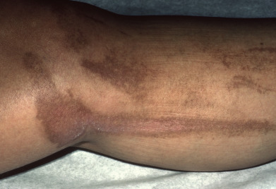
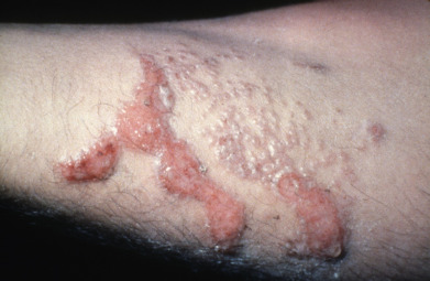
Cutaneous larva migrans demonstrates an erythematous serpiginous lesion. The worm is located ahead of the advancing border of the lesion, as the cutaneous reaction is a manifestation of a delayed-type immune response to the organism.


Recognition of arthropods of medical importance is critical to allow assessment of the risk of vector-borne disease and guide management. The skin is often affected by ectoparasites as well as endoparasites, and this section of the atlas will provide a guide to identification of the most important organisms.


Fig. 20.1


Old World leishmaniasis.


Fig. 20.2


Old World leishmaniasis.


Fig. 20.3


Old World leishmaniasis.


Fig. 20.4


New World leishmaniasis.


Fig. 20.5


New World leishmaniasis.


Fig. 20.6


New World leishmaniasis.


Fig. 20.7


New World leishmaniasis.


Fig. 20.8


New World leishmaniasis.

Courtesy Scott Norton, MD.


Fig. 20.9


New World leishmaniasis.


Fig. 20.10


New World leishmaniasis.


Fig. 20.11


New World leishmaniasis.


Fig. 20.12


Mucocutaneous leishmaniasis, same patient as Fig. 20.11 .


Fig. 20.13


Mucocutaneous leishmaniasis.


Fig. 20.14


Mucocutaneous leishmaniasis.


Fig. 20.15


Mucocutaneous leishmaniasis.

Courtesy Lauro de Souza Lima Institute, Brazil.


Fig. 20.16


Anergic leishmaniasis.


Fig. 20.17


Anergic leishmaniasis.

Courtesy Lauro de Souza Lima Institute, Brazil.


Fig. 20.18


Anergic leishmaniasis.


Fig. 20.19


Post-kala-azar dermal leishmaniasis.

Courtesy Debabrata Bandyopadhyay, MD.


Fig. 20.20


Triatome bite.


Fig. 20.21


Romana sign, Chagas disease.


Fig. 20.22


Toxoplasmosis. Note erythema of eyelids.

Courtesy Steven Binnick, MD.


Fig. 20.23


Portuguese man of war dermatitis.

Courtesy Rui Tavares Bello, MD.


Fig. 20.24


Jellyfish dermatitis.

Courtesy Steven Binnick, MD.


Fig. 20.25


Sea wasp dermatitis.

Courtesy Curt Samlaska, MD.


Fig. 20.26


Healed jellyfish dermatitis.

Courtesy Steven Binnick, MD.


Fig. 20.27


Seabather eruption.

Courtesy Scott Norton, MD.


Fig. 20.28


Fire coral dermatitis.

Courtesy Steven Binnick, MD.


Fig. 20.29


Coral granuloma.


Fig. 20.30


Sea urchin injury.

Courtesy Scott Norton, MD.


Sep 3, 2019 | Posted by in Dermatology | Comments Off on Parasitic Infestations, Stings, and Bites

Full access? Get Clinical Tree

Get Clinical Tree app for offline access