| Malignant primary orbital tumors |
| Malignant eyelid and ocular adnexal tumors with orbital extension |
| Malignant orbital tumors extending from the cranium or paranasal sinuses |
| Malignant intraocular tumors with extrascleral extension |
| Sino-orbital invasive fungal infections |
| Orbital metastatic disease or advanced orbital disease as palliative therapy |
| Definitive pathologic diagnosis of malignancy |
| Systemic oncologic workup to include orbitofacial imaging |
| Neurosensory examination (ophthalmic V1 and maxillary V2) |
| Lymph node examination |
| Discussion of expectations of surgery, rehabilitation, psychological impact |
| Sinonasal evaluation if extension into sinuses or fungal etiology |
Introduction
Orbital exenteration is an operation that must be reserved for life-threatening or severely progressive disease that is not amenable to alternative treatment. It is a disfiguring operation that can extend survival, relieve pain and improve appearance in certain circumstances but should not be contemplated without considerable deliberation and extensive discussion with the patient and family.
Common indications for exenteration include primary orbital malignancy, orbital extension of adnexal tumors (including skin cancers and sinus tumors), extrascleral extension of primary ocular tumors, intractable pain, life-threatening infection, and extensive ocular surface malignancy. Orbital exenteration can be surgically tailored in response to the indication with more extensive orbital tissue removal being desired when surgical cure is the goal and more limited removal when palliation or pain control is desired.
Multidisciplinary tumor board involvement is critical in the management of complex orbital malignancies. Colleagues with expertise in radiation and chemotherapy should be readily available for consultation as needed during adjunct therapy. Sometimes radical surgery is not the best option and input from other services is essential to help manage difficult tumor cases.
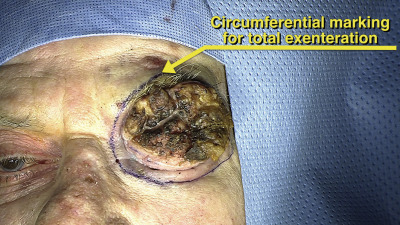
There are different types of exenteration. Total exenteration, involves removal of all orbital tissue including the eyelids, globe, orbital soft tissues and periorbita. Adjacent bone and the sinuses may also be included in the removal. A limited or subtotal exenteration involves globe removal with the sparing of some orbital soft tissue, limiting the excision to the anterior orbit. Either total or subtotal exenteration may be combined with sparing of the eyelids, which allows for faster healing and results in less disfigurement.
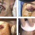
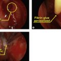
Reconstruction of the exenterated socket can be accomplished in several ways. Secondary intention with granulation typically takes weeks and involves frequent dressing changes and prolonged discomfort. Split-thickness skin grafts allow for more rapid healing of the socket but harvesting of skin must be from a secondary donor site. If eyelid skin can be preserved, the residual skin can be used to line the orbit for the most rapid healing. A myocutaneous free flap can be used if additional volume is desired, but this typically precludes use of an exenteration prosthesis and may obscure tumor recurrence. An additional option includes the use of a vascularized temporalis flap that is tunneled through the lateral orbital rim and can be used to support a variety of grafts, including autologous dermis fat.
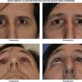
Provided there is adequate space, good cosmesis can be achieved with a custom orbital exenteration prosthesis that includes a globe and non-functioning eyelids. Large frame glasses can be fitted to help camouflage the prosthetic rim. Osseointegration with retaining posts can be implanted into the orbit to facilitate retention of a prosthesis with a difficult-to-fit orbit. Alternatively a patch can be used.
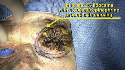
Surgical Technique