3 Orbit and Facial Bone
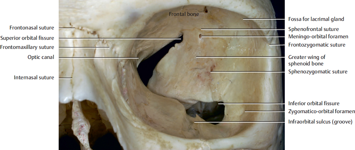
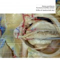
Fig. 3.1. Anterior view of the orbit.
The inferior orbital fissure lies at the junction of the lateral wall and the floor of the orbit. Through this fissure pass the infraorbital and zygomatic branches of the maxillary division of the trigeminal nerve and accompanying vessels.
The infraorbital nerve is the terminal branch of the maxillary nerve. It courses along the orbital floor in the infraorbital sulcus (groove) into a canal and onto the face at the infraorbital foramen.
The zygomatic nerve arises from the maxillary nerve in the pterygopalatine fossa and passes through the inferior orbital fissure to course along the lateral wall of the orbit, where it divides into zygomaticofacial and zygomaticotemporal branches. The branches enter the zygomatico-orbital foramina on the orbital surface of the zygomatic bone. When one foramen is present, the zygomatic vessels and nerve enter and then branch within the bone to exit different foramina as the zygomaticofacial and zygomaticotemporal vessels and nerve. The zygomaticotemporal nerve provides sensation to the temporal skin, and the zygomaticofacial nerve provides sensation to the skin over the prominence of the cheek.
Meningo-orbital foramen (lacrimal foramen) is located in the greater wing of the sphenoid, anterior to the tip of the superior orbital fissure, and is the source of an anastomosis between the lacrimal artery and the orbital branch of the middle meningeal artery. This foramen is present in approximately 50% of Caucasian people.
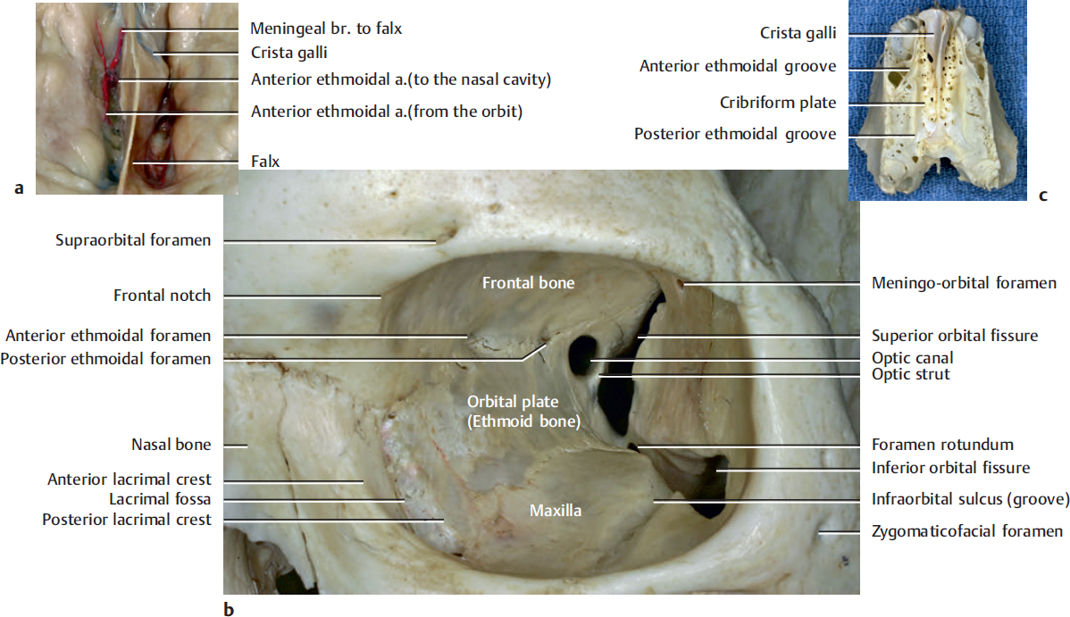
The supraorbital foramen, the frequency of which is 20 to 30%, is the passage mainly for the supraorbital nerve, especially its lateral branch, one of the medial branches, and its concomitant small artery (see Fig. 5.3b). The main trunk of the supraorbital artery generally passes under the supraorbital notch or just below the supraorbital rim. The supraorbital notch also transmits the supraorbital nerve. The frontal notch, which is medial to the supraorbital notch, transmits the supratrochlear nerve and artery.
The optic canal lies within the lesser wing of the sphenoid and transmits the optic nerve and ophthalmic artery.
The anterior and posterior ethmoidal foramina for the same nerves and vessels are situated at the medial wall. The anterior ethmoidal nerve exits the orbit through the anterior ethmoidal foramen and enters the anterior cranial fossa. It runs into the roof of the nose through small slits lying on each side of the crista galli. The posterior ethmoidal nerve exits the orbit through the posterior ethmoidal foramen to enter the nose. It supplies the sphenoidal sinus and the posterior ethmoidal air cells. The view of the ethmoid bone from above (Fig. 3.2c) shows anterior and posterior ethmoidal grooves for the same nerves and vessels, which are converted into foramina by articulation with frontal bone. It is important that lowest point of the cribriform plate (lowest point of the anterior skull base) is generally below the anterior and posterior ethmoidal foramen on the medial orbital wall (Fig. 3.2a).
The fossa for the lacrimal sac is formed by the lacrimal incisure on the maxilla and a matching groove on the lacrimal bone. When the adjacent grooves are combined, they form a fossa and a canal that houses the lacrimal sac and transmits the beginning of the lacrimal duct toward the nasal cavity.
Related posts:

Stay updated, free articles. Join our Telegram channel

Full access? Get Clinical Tree


