11 Lower Facial Region
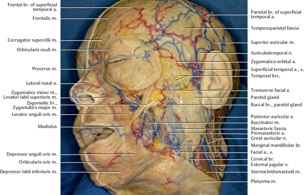
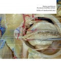
Fig. 11.1. Lateral oblique view of the face. The skin has been removed.
The modiolus is a nodular region at the corner of the mouth where several mimetic muscles converge and interlace. It is contributed by the orbicularis oris, buccinator, levator anguli oris, depressor anguli oris, zygomatics major, and risorius muscles.
The depressor labii inferioris muscle is partly covered by the anterior fibers of the depressor anguli oris muscle.
The levator anguli oris, the buccinator, and the mentalis muscles are deep-seated muscles that are innervated on their superficial surface by the facial nerve branches.
The facial vein begins as the angular vein at the medial corner of the eye. The angular vein is formed by the confluence of the supraorbital and supratrochlear veins. The facial vein passes downward behind the facial artery independently to the inferior border of the mandible.
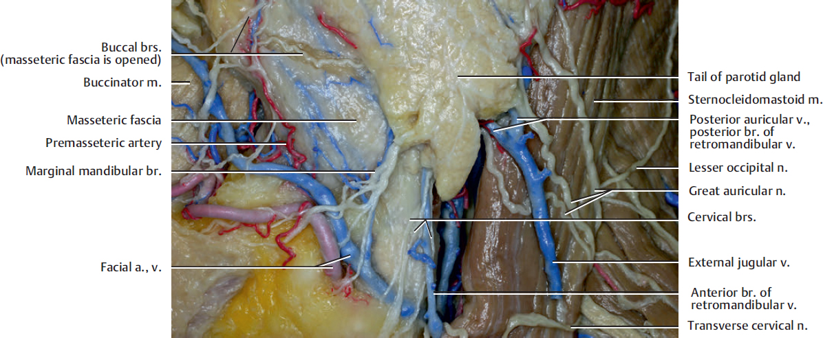
The marginal mandibular branches of the facial nerve emerge from the lower border of the parotid gland, and the branch runs near the inferior border of the mandible. Initially, it may pass into the neck below the angle of the mandible, but after crossing the facial artery it usually comes up above the lower border of the mandible to the face. It innervates the depressor anguli oris, the depressor labii inferioris, the mentalis, and the lower orbicularis oris muscles.
The cervical branch passes downward from the lower border of the parotid gland to supply the platysma muscle in the neck. Some of the its branches frequently innervate the depressor labii inferioris and the depressor anguli oris muscles.
One of the buccal branches runs beneath the masseteric fascia in this figure. However, most of the marginal mandibular branches generally run on the fascia.
At the inferior border of the mandible near the anterior edge of the masseter muscle, the facial artery and the vein course closely.
The premasseteric artery arises at the lower border of the mandible from the facial artery and ascends along the anterior edge of the masseter muscle.
The great auricular nerve perforates the deep cervical fascia and ascends upon the sternocleidomastoid muscle beneath the platysma to the parotid gland, where it divides into an anterior and a posterior branch. It provides sensory innervation for the skin over the parotid gland and the mastoid process, as well as both surfaces of the caudal region of the auricle.
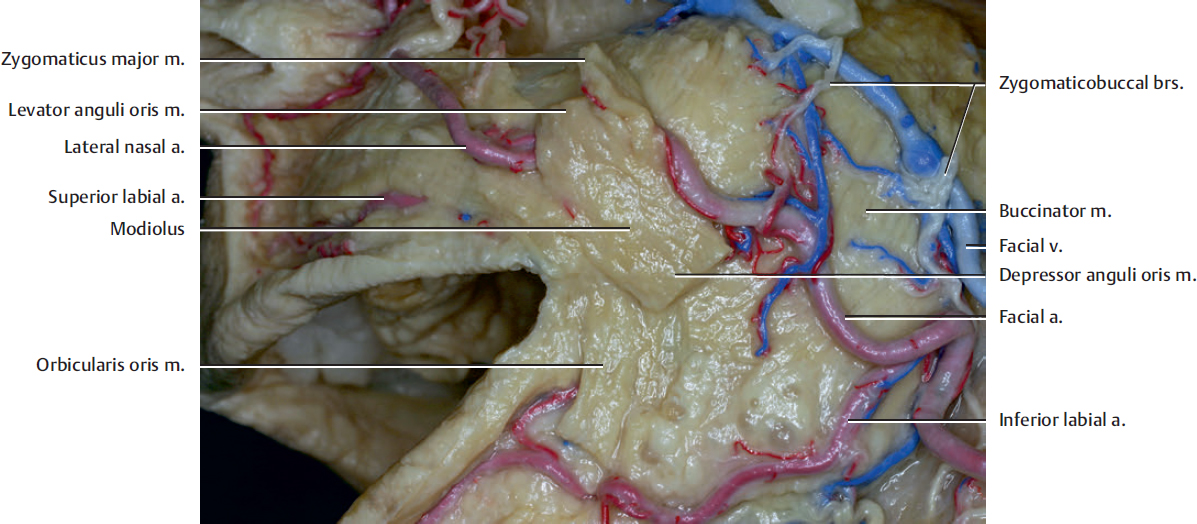
The modiolus may be fixed by the action of some of the muscles to provide a base for the action of other muscles.
Related posts:

Stay updated, free articles. Join our Telegram channel

Full access? Get Clinical Tree


