7 Superficial Structures in the Midfacial Region
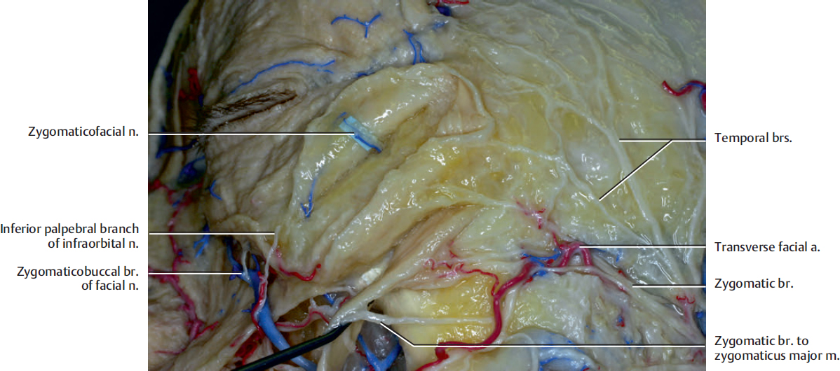
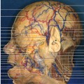
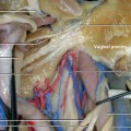
Fig. 7.1. The midfacial region. The lower lateral portion of the orbicularis oculi muscle has been reflected.
Innervation to the lower orbicularis oculi muscle and zygomaticus major muscle are shown. The zygomaticus major muscle is innervated by the zygomatic branches and also from the zygomaticobuccal branches (see Fig. 7.5) in its lower part of the muscle. The lower orbicularis muscle is innervated from its inferolateral side by one or two zygomatic branches and temporal branches.
The zygomaticofacial nerve (shown in Fig. 7.1 with blue sheet under it) is a sensory nerve, which supplies the small region of the cheek, and it is divided from the zygomatic nerve in the orbit.
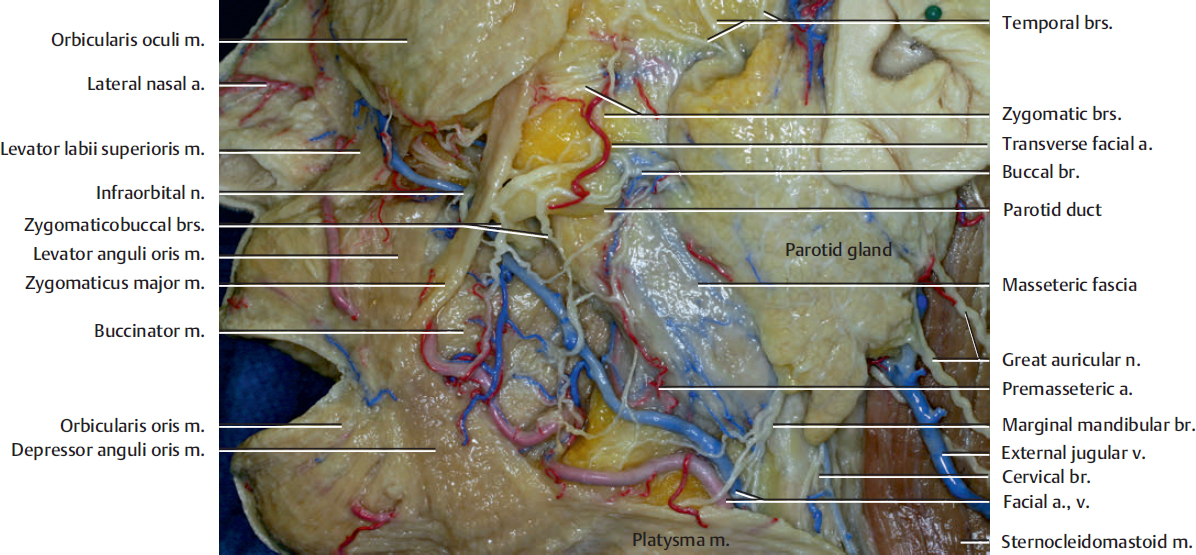
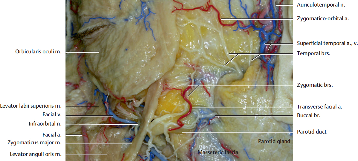
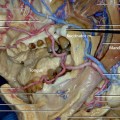
The transverse facial artery, which originates from the superficial temporal artery within the parotid gland, courses anteriorly beneath the masseteric fascia above the parotid duct.
The buccinator muscle, upper orbicularis oris muscle, and levator anguli oris muscle are innervated by the zygomaticobuccal branches (see Fig. 7.5). The buccinator muscle and the levator anguli oris muscle are the deep-seated muscles and they are innervated from their superficial surface.
The anterior margin of the parotid gland is a site which is suitable to identify the zygomatic and buccal branches for facial reanimation surgery. Several zygomatic and buccal branches can be exposed via the small skin incision made just below the zygomatic arch and anterior to the mandibular notch. The notch can be identified by palpation. These branches can also be exposed via preauricular skin incision. The careful selection of the donor facial nerve branches is mandatory for facial reanimation surgery because some of the buccal and zygomaticobuccal branches might not innervate the zygomaticus major muscle, which is the target muscle for smile reconstruction.
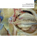
The levator anguli oris muscle arises from the canine fossa of the maxilla, immediately below the infraorbital foramen, and passes downward toward the corner of the mouth. It elevates the corner of the mouth.
The facial nerve branches do not always run on the same plane. Some of the zygomatic branches run beneath the masseteric fascia anterior to the parotid gland. It is also an important surgical point that we can only divide the parotid gland just over the facial nerve branch during the dissection of the facial nerve branches in the parotid gland.
The buccal branch of the facial nerve has a close relationship with the parotid duct. The branch is most likely inferior and within 1 cm to the duct.
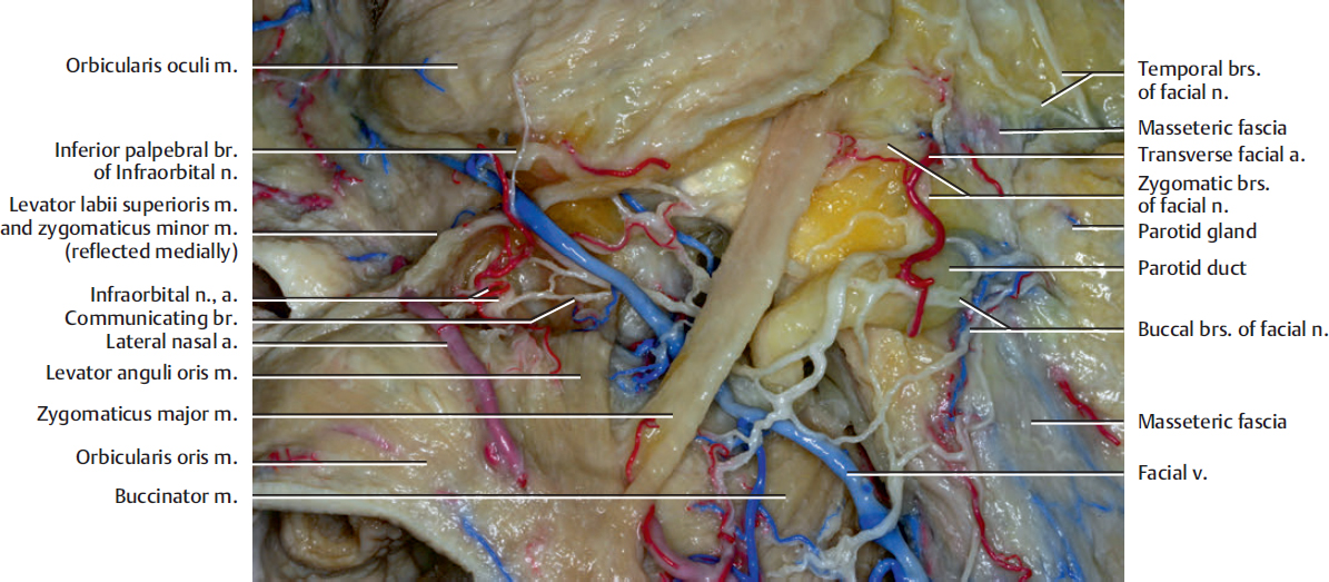
Some of the zygomaticobuccal branches (see Fig. 7.5) course downward along the parotid duct, pass under the zygomatic major muscle, then course upward to reach the lower orbicularis oculi muscle, and finally end up at the medial canthal region. The upper orbicularis oris muscle is generally innervated by the zygomaticobuccal branches. The branches reach its upper lateral side, which is medial to the zygomaticus major muscle.
The levator anguli oris and buccinator muscles are in the deepest layer of the mimetic muscles and are innervated from their superficial surface. The levator anguli oris muscle, which originated from the canine fossa, inserts into the modiolus.
The facial nerve branches have communications with the branches of the infraorbital nerve.
Related posts:

Stay updated, free articles. Join our Telegram channel

Full access? Get Clinical Tree


