10 Deep Structures in the Midfacial Region
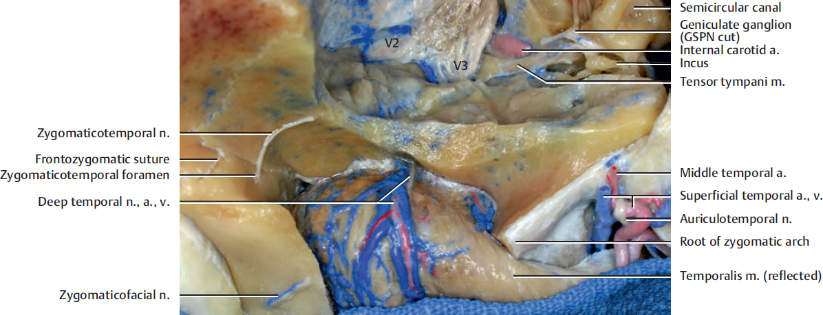
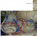
Fig. 10.1. Superior view of the middle fossa and temporal fossa.
The neurovascular bundle is shown on the deep surface of the temporalis muscle. The dissection of the muscle from the temporal bone might cause the muscle to atrophy because of damage to its motor nerves.
The zygomaticotemporal nerve is a terminal branch of the maxillary division of the trigeminal nerve. The zygomatic nerve enters the orbit through the inferior orbital fissure, and it divides into the zygomaticotemporal and zygomaticofacial nerves along the lateral wall of the orbit. The zygomaticotemporal nerve emerges from the orbit into the temporal fossa at approximately 14 mm inferior to the frontozygomatic suture and 10 mm lateral to the lateral margin of the orbit through the zygomaticotemporal foramen. Then, it passes through the temporalis muscle and pierces the temporal fascia about 2 cm above the zygomatic arch. The foramen is always found posterolateral to the edge of the lateral orbital rim. It is a sensory nerve that provides sensation to the skin of the temporal region.
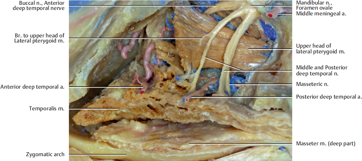
The buccal nerve and the anterior deep temporal nerve generally pass between the upper and lower heads of the lateral pterygoid muscle. The middle and posterior deep temporal nerve and masseteric nerve run on the superior surface of the upper head.
The temporalis muscle is innervated predominantly by the deep temporal nerves, which are branches of the anterior trunk of the mandibular nerve. The number of the deep temporal nerves generally vary from one to five. The temporal branches, which innervate the temporalis muscle, also arise from the buccal nerve and the masseteric nerve. They innervate the anterior and posterior part of the temporalis muscle respectively.
Three main arteries supply the temporalis muscle: the anterior deep temporal, the posterior deep temporal, and the middle temporal. The anterior deep temporal artery, which arises from the pterygoid segment of the internal maxillary artery, enters the anterior portion of the muscle and supplies about 30% of the muscle. The posterior deep temporal artery, which arises from the same segment of the maxillary artery, enters the central part of the muscle and supplies about 50% of the muscle. The middle temporal artery. which arises from the superficial temporal artery, supplies the temporal fascia and approximately 20% of the posterior and upper parts of the temporalis muscle.
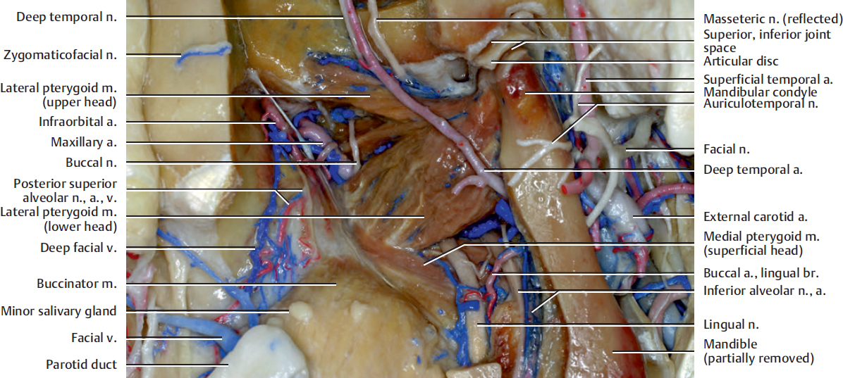
The lingual nerve is a branch of the posterior trunk of the mandibular nerve. It is essentially a sensory nerve. It emerges from the inferior border of the lateral pterygoid muscle and curves downward and forward in the space between the ramus of the mandible and the medial pterygoid muscle.
The maxillary artery is a terminal branch of the external carotid artery. It arises at the posterior border of the mandibular neck. It is divided into three segments. The first is the mandibular segment, which lies behind the mandible and courses horizontally between the neck of the mandible and lateral to the sphenomandibular ligament. This segment has five branches and all enter bone, where the segment lies parallel to and a little below the auriculotemporal nerve. It crosses the inferior alveolar nerve and runs along the lower border of the lateral pterygoid muscle. These branches include the deep auricular artery, the anterior tympanic artery, the middle meningeal artery, an accessory meningeal artery, and the inferior alveolar artery.
The inferior alveolar artery and vein descend downward and forward to join the inferior alveolar nerve. They descend to the mandibular foramen on the medial surface of the ramus of the mandible and run along the mandibular canal in the substance of the bone. Opposite the first premolar tooth, they divide into two branches: incisor and mental.
The temporomandibular joint (TMJ) is formed by the condyle of the mandible articulating in the mandibular fossa (glenoid fossa) of the temporal bone. The joint cavity is divided into two by an intra-articular disc. Its margins merge with the joint capsule. The TMJ is basically a hinge joint, but it also allows for some gliding movements. Sensory innervation of the temporomandibular joint is derived from the auriculotemporal nerve and the articular branch from the masseteric nerve.
The lateral pterygoid muscle runs in the horizontal plane and occupies most of the infratemporal fossa. It has two distinct heads: a smaller upper infratemporal head and a lower pterygoid one. The infratemporal head originates at the lateral surface of the greater sphenoid wing and the infratemporal crest of the sphenoid bone. It runs parallel to the floor of the middle cranial fossa and merges posteriorly with the pterygoid head. The pterygoid head originates from the lateral surface of the lateral pterygoid plate and runs laterally and superiorly. Both heads were inserted in a depression at the anterior aspect of the neck of the mandible. The muscle assists in opening the jaws by pulling forward the mandibular condyle and the articular disc of the temporomandibular joint. The muscle is innervated by the mandibular nerve.
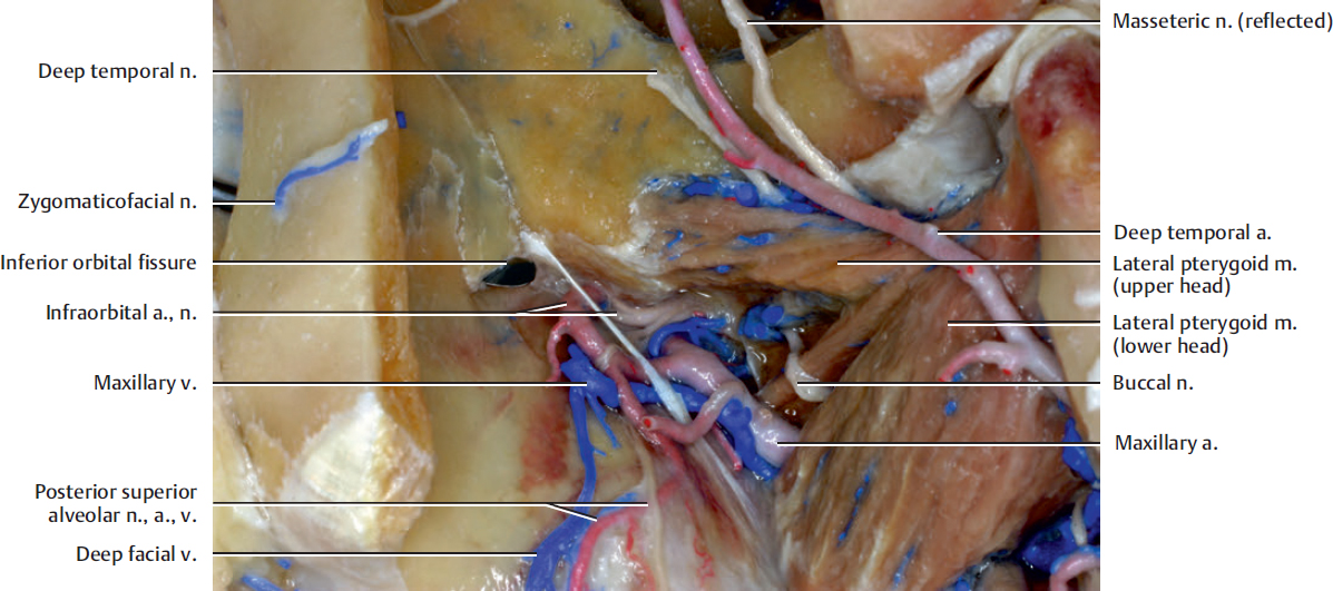
The posterior superior alveolar artery arises from the maxillary artery (pterygopalatine segment), frequently in conjunction with the infraorbital artery just as the maxillary artery is passing into the pterygopalatine fossa. It runs onto the maxillary tuberosity and supplies the maxillary molar and premolar teeth through the alveolar canal (see Fig. 10.13b), the buccal gingiva, and the maxillary air sinus. The posterior superior alveolar vein drains into the pterygoid venous plexus or the maxillary vein.
The inferior orbital fissure transmits the maxillary nerve and its zygomatic branch, the infraorbital vessels, the ascending branches from the pterygopalatine ganglion, and a vein that connects the inferior ophthalmic vein with the pterygoid venous plexus. The orbitalis muscle (Müller’s muscle) occupies the lateral part of the inferior orbital fissure and forms a lamina of smooth muscle fibers that cover the inferior orbital fissure. The orbitalis muscle is a rudimentary smooth muscle that crosses from the infraorbital groove and sphenomaxillary fissure and is intimately united with the periosteum of the orbit. It lies at the back of the orbit and spans the infraorbital fissure.
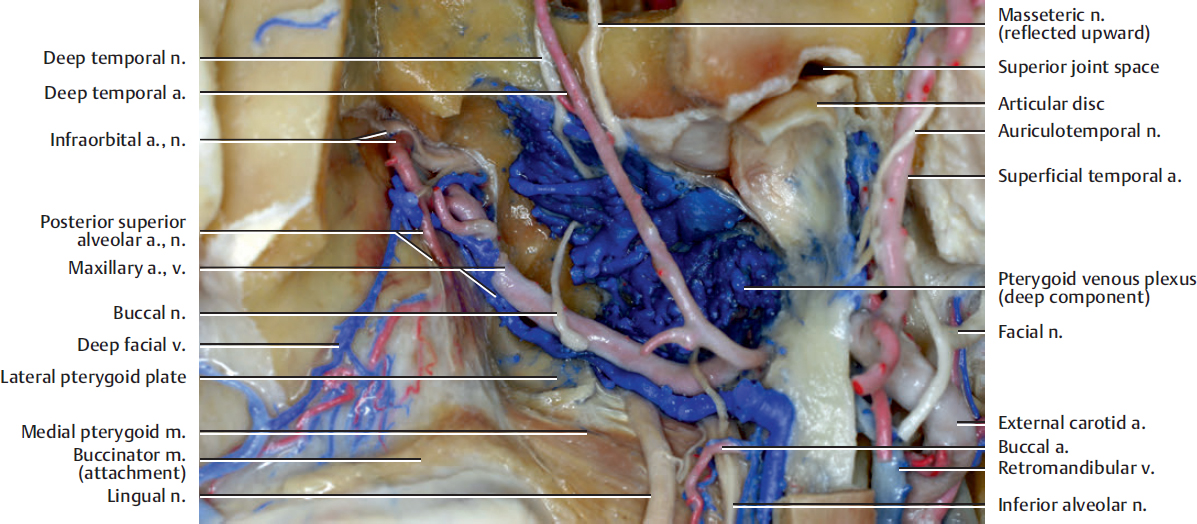
The medial pterygoid muscle originates in the pterygoid fossa from the medial surface of the lateral pterygoid plate. Its fibers pass inferiorly to attach to the medial surface of the ramus and angle of the mandible. It elevates the mandible.
The pterygoid segment, which is the second segment of the maxillary artery, is related to the pterygoid head of the lateral pterygoid muscle. It runs between the infratemporal and pterygoid heads of the lateral pterygoid muscle or laterally to the pterygoid head of the lateral pterygoid muscle. It has five branches: the deep temporal arteries (anterior and posterior), the pterygoid artery, the masseteric artery, the buccal artery, and a small lingual branch.
The pterygoid venous plexus has superficial and deep components. The latter is more prominent and is located between the lateral and medial pterygoid muscles, posterior to the lateral pterygoid plate, around the lingual and inferior alveolar nerves. It communicates with the cavernous sinus, the facial vein, the retromandibular vein, the inferior ophthalmic vein, and the pharyngeal plexus. The primary drainage of the pterygoid venous plexus is posteriorly through the retromandibular vein via the maxillary vein. The deep facial vein connects the facial vein with the pterygoid venous plexus in the infratemporal fossa.
Related posts:

Stay updated, free articles. Join our Telegram channel

Full access? Get Clinical Tree


