5 Forehead and Orbital Region
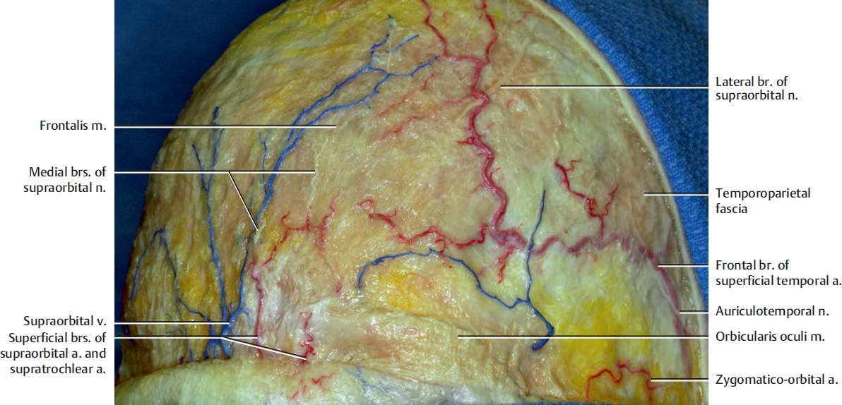
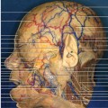
Fig. 5.1. The frontotemporal region. The skin has been removed to show the frontalis muscle and galeal layer.
The galea aponeurotica extends from the external occipital protuberance and supreme nuchal lines to the eyebrows. The aponeurotica is continuous laterally with the temporoparietal fascia overlying the temporal fascia. The galea aponeurotica contains the occipitofrontal muscle. Each frontal belly of the frontalis muscle arises from the anterior margin of the galea aponeurotica and passes forward to merge with the orbicularis oculi muscle. The main function of the occipitofrontalis muscle is to elevate the eyebrows to produce transverse furrows of the forehead. This frontalis muscle is innervated by the temporal branch of the facial nerve.
The forehead sensation is supplied by the supraorbital and supratrochlear nerves. The supratrochlear nerve provides sensation to the medial side of the forehead. The supraorbital nerve has medial (superficial) and lateral (deep) branches. The former provides sensation to the forehead region, and the latter supplies sensation to the frontoparietal region. The lateral branch penetrates the frontalis muscle and galeal layer at the forehead, usually above the hairline.
The superficial temporal artery and vein run on the galeal aponeurotica. The superficial branches of the supraorbital and supratrochlear arteries communicate with the superficial temporal artery on the galeal layer.
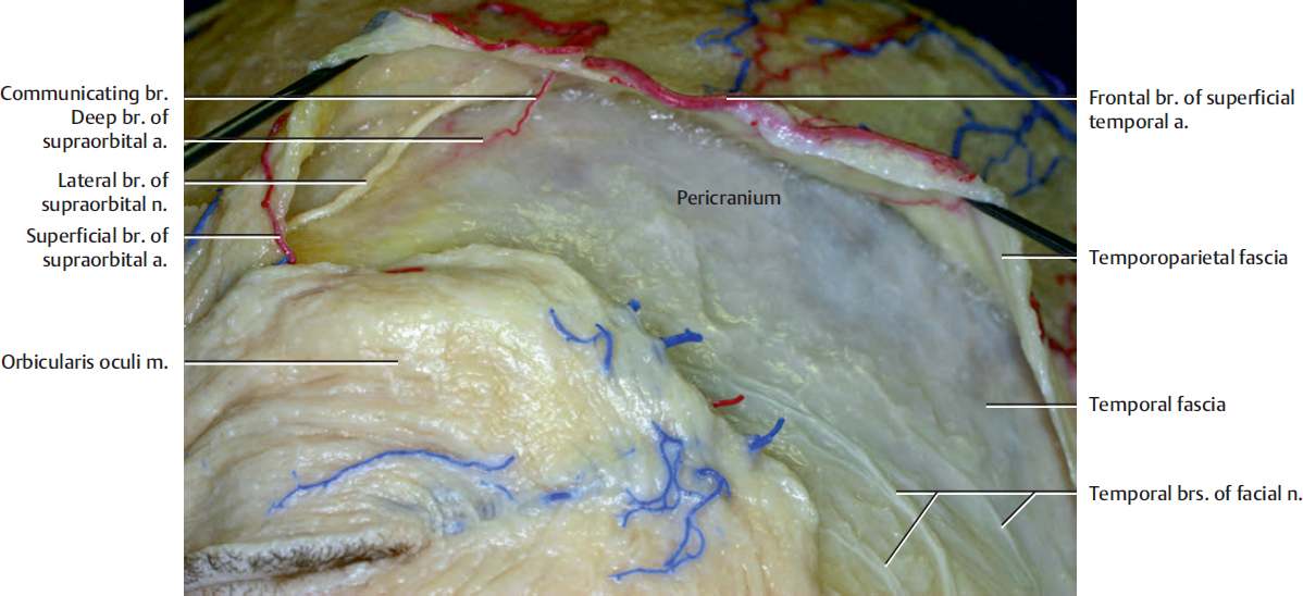
The temporal branches of the facial nerve generally course along the undersurface of the temporoparietal fascia and in the subgaleal fat pad. The temporoparietal fascia (galea) and its extension frontalis muscle are elevated to leave the temporal branches of the facial nerve on the temporal fascia in this specimen.
The deep (lateral) division of the supraorbital nerve runs cephalad across the lateral forehead between the frontalis muscle and galeal layer and the pericranium as the sensory nerve to the frontoparietal scalp. There is a loose areolar tissue between the galea aponeurotica and the pericranium that allows scalp mobility.
The arterial communication between the superficial temporal artery and the deep branch of the supraorbital artery is shown.
The pericranium is continuous with the temporal fascia in the temporal region.
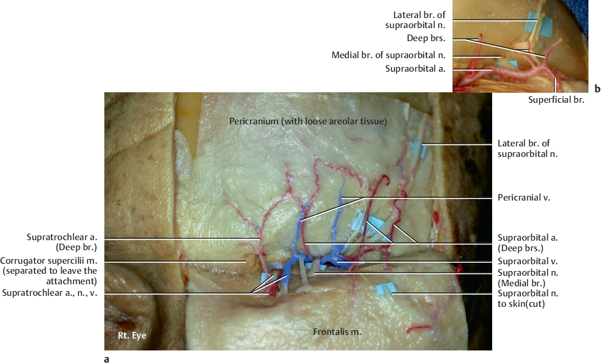
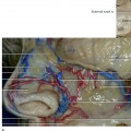
The frontalis muscle has been reflected inferiorly. The attachment of the corrugator supercilii muscle is shown at the medial side on the frontal bone.
The supraorbital nerve has two divisions: a superficial (medial) division that passes shortly over the pericranium and then pierces the frontalis muscle, providing sensory supply to the forehead skin, and a deep (lateral) division that runs cephalad across the lateral forehead between the galea aponeurotica and the pericranium as the sensory nerve to the frontoparietal scalp. The deep division consistently courses approximately 1 cm medial to the superior temporal line, which is the attachment of the temporal fascia. It can be identified at the level of hair line just beneath the galea aponeurotica. In contrast, the supratrochlear nerve has only a superficial branch.
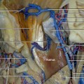
The main trunk of the supraorbital and supratrochlear arteries course below the orbital roof and divide near or above the supraorbital rim into superficial and deep branches (Fig. 5.3b). The superficial branch run in the galea-frontalis layer of the scalp, and the deep branches ascend in and supply the pericranium. The deep branch of the supratrochlear artery generally penetrates the corrugator supercilii muscle before reaching the pericranium. The forehead pericranium is supplied dominantly from the deep branches of the supraorbital artery.
Both the deep veins from the pericranial layer and the superficial veins from the galea-frontalis layer empty into a transverse channel in the supraorbital area that courses between the galea-frontalis layer and the pericranium. This transverse venous trunk joins the supratrochlear veins on the medial side and the superficial temporal veins on the lateral side.
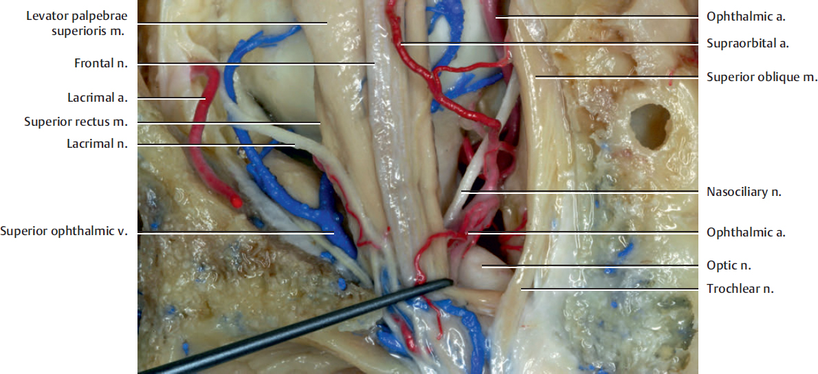
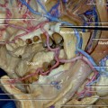
The ophthalmic nerve passes along the lateral dural wall of the cavernous sinus and gives off three main branches just before the superior orbital fissure. The three branches are the lacrimal nerve, the frontal nerve, and the nasociliary nerve.
Related posts:

Stay updated, free articles. Join our Telegram channel

Full access? Get Clinical Tree


