Fig. 15.1
(a) Optical coherence tomography (OCT) image of normal skin taken just adjacent to an actinic keratosis (AK) lesion located on the scalp. White arrows mark the dermoepidermal junction (DEJ). Black arrows mark vessels. (b) OCT image of AK lesion located on the scalp. Vertical white lines mark disruption of layering. Thick black arrow marks characteristic white streak in the epidermis. White arrows mark the DEJ
15.3 Optical Coherence Tomography Monitoring of Cryotherapy Treatment
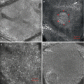
OCT imaging of noninvasive treatment modalities has recently been investigated while OCT imaging in connection with invasive treatments like simple excisions and Mohs surgery has been more extensively studied [12, 14–17]. Cryosurgery of premalignant lesions like AK is traditionally guided clinically by freeze-thaw time and lateral extent of freezing, and therefore, real-time OCT monitoring of cryo treatment has been thought to potentially improve accuracy and efficacy of the procedure. Unfortunately, the immediate effects of cryotherapy impair OCT imaging of the tissue. Cryotherapy induces an opaque iceball in the tissue, which completely prevents visualization of the treated lesion and the freezing depth [18] (Fig. 15.2a–d). The reason for the opacity is that at the OCT wavelength of 1,300 nm, ice is around ten times more absorbing than liquid water. When the tissue freezes the created ice absorbs all of the OCT laser light, and therefore, OCT images of cryotherapy-treated skin only show an impervious frosty white line at the skin surface. Normal OCT imaging depth and resolution reappears in time with the thawing of the tissue. In cryosurgery-treated grade 1 and 2 AK lesions, OCT imaging has showed vesicle formation in the lesions within twenty minutes after treatment [18]. The vesicles were primarily located along the dermoepidermal junction and appeared as dark areas with a maximum height of 0.47 mm (Fig. 15.2a–d). The vesicles were subclinical by the time of the post-cryosurgery imaging and could thus only be identified using OCT. A vesiculobullous reaction after superficial cryogen application is an indication of successful treatment of superficial epidermal lesions because the separation of epidermis and dermis enables the shedding of the diseased skin [19].
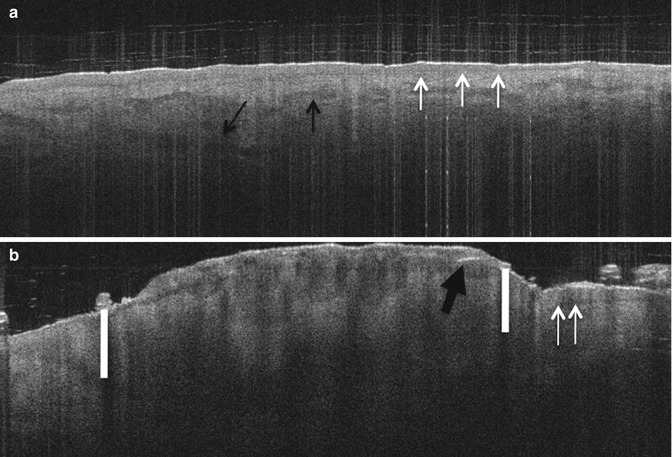
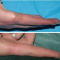
Fig. 15.2
(a) Optical coherence tomography (OCT) image of normal skin taken just adjacent to an actinic keratosis (AK) lesion located on the dorsal side of a hand. White arrows mark the dermoepidermal junction (DEJ). Black arrows mark vessels. (b) OCT image of AK lesion before cryo treatment. Vertical white lines mark disruption of layering caused by thickening of the epidermis. Thick black arrows mark characteristic white streaks in the epidermis. Thin black arrow marks a hair casting a shadow. (c) OCT image taken seconds after cryosurgery. The treatment produces an opaque iceball. (d) OCT image taken 20 min after cryotherapy. Stars mark emerging vesicles along the DEJ. White arrows mark the DEJ. Small black arrow marks a hair casting a shadow
15.4 Conclusions
The capability of OCT to identify early tissue changes after cryosurgery of AK lesions implies that OCT could potentially be used as a supplementary tool in assessing the effectiveness of cryosurgery, but specific follow-up studies have not yet been performed. Results from a study on OCT imaging of noninvasive photodynamic therapy of keratinocyte carcinomas and AK indicate that subclinical residual lesions could be identified by OCT at 3 months follow-up [13]. OCT may have the potential to influence patient management by enabling early evaluation of treatment effects. Early detection of complete or partial responses to invasive and noninvasive therapies could make for a better planning of treatment, e.g., reducing treatment area, number of treatments, and thereby reducing pain related to re-treatment. In regard to imaging of the skin and monitoring of treatments, OCT is a relatively novel technology and the cryosurgery and follow-up results are based on studies with small sample sizes. In our view the results are promising and the technology has achieved a level of maturation that makes it possible to explore its clinical use on a larger scale. Additional studies focusing on both the use of OCT as a tool for treatment planning and for follow-up after skin cancer treatments are warranted before OCT can be established as an important tool in the daily clinical practice, but until now more than 15 clinical studies imply that OCT imaging does have a potential as a diagnostic imaging tool in dermatology [8, 20, 21].
Essential Tidbits
OCT is an imaging tool that provides real-time high-resolution skin images to a depth of up to 2 mm.
It has been used in monitoring cryosurgery of AK.
OCT imaging has shown subclinical vesicle formation in the lesions within twenty minutes after cryosurgical AK treatment.Related posts:
 Preoperative Care for Cryosurgery
 Theoretical Principles of Immunocryosurgery
 Cryobiopsy, Cryoanesthesia, and Cryoanalgesia
 In Vivo Reflectance Confocal Microscopy Assessment of Wound Induction and Repair of a Skin Injury Produced by Liquid Nitrogen: An Atlas
 Cryosurgery for Warts
 Cryosurgery for Vascular Lesions

Stay updated, free articles. Join our Telegram channel

Full access? Get Clinical Tree


