34 Optic Nerve Sheath Fenestration
Idiopathic intracranial hypertension (IIH) is a syndrome characterized by increased intracranial pressure (ICP) without clinical, laboratory, or radiological evidence of a space-occupying lesion, hydrocephalus, or meningitis. The modified Dandy’s criteria for this condition are signs and symptoms of increased ICP (headaches, nausea, vomiting, transient visual obscuration, papilledema), awake and alert patient, no localizing neurological signs other than unilateral or bilateral abducens nerve paresis, documented increased CSF pressure (>200 mm of H2O in nonobese and >250 mm of H2O in obese) with normal CSF composition, normal neuroimaging studies except for small ventricles or empty sella, and no other cause of intracranial hypertension present. Although pseudotumor cerebri occurs in both sexes, from childhood to the seventh decade, most patients are obese women of child-bearing age.
The pathophysiological basis of IIH remains unclear, but possible explanations for IIH include increased CSF production, sustained increase in intracranial venous pressure, increased resistance to CSF absorption by the pacchionian granulation, and increase in brain volume because of an increase in the cerebral blood volume or interstitial fluid volume simulating a form of brain edema. Increased venous sinus pressure is now hypothesized to be the putative cause of IIH.
The major complication of this disorder is visual loss, occurring in 10% to 26% of patients due to nerve fiber damage at the optic disc caused by papilledema. The natural history of patients with IIH followed over a long period demonstrates that around 50% will have some permanent visual deficit and severe visual field defects in 25%. It is essential that visual functions be monitored carefully in all patients with papilledema, as optic disc appearance alone may be misleading.
Medical therapy is directed at preservation of vision and relief of headaches. The drugs of choice are carbonic anhydrase inhibitors (CAIs) such as acetazolamide, or a loop diuretic such as furosemide to depress CSF production. In cases with significant visual loss, intravenous acetazolamide and corticosteroids can be utilized acutely for a short time.
If visual loss progresses despite optimum medical treatment, surgical decompression should be considered sooner rather than later, before major visual field or acuity deficit supervenes.
Bilateral subtemporal decompression, although effective, is rarely performed because of a high rate of serious complications. Lumbar-peritoneal shunts, ventriculoperitoneal shunts, and cisternal-peritoneal shunts have been used, but the risks of infection or shunt blockage have limited their acceptance. Optic nerve sheath fenestration is a well-accepted surgical technique employed to preserve vision in patients with papilledema that threatens vision. Preoperative screening for metabolic or electrolyte abnormalities must be performed in patients who have been receiving long-term medical therapy, especially CAIs or diuretics. Systemic hypertension may contribute to optic disc damage and visual morbidity in these patients. Although systemic hypertension should be treated, abrupt or drastic reduction of blood pressure must be avoided since it may result in hypoperfusion to the optic disc in the presence of marked disc edema.
Access to the optic nerve sheath can be achieved through a transconjunctival medial orbitotomy, lateral orbitotomy, or superomedial lid crease approach. The lateral approach is useful for patients with failed medial optic nerve sheath fenestration or in patients with previous scleral buckles that limit optic nerve exposure.
All optic nerve sheath fenestrations are performed under general anesthesia, as optimal patient cooperation is essential. While under general anesthesia, the patient is placed in a slight reverse Trendelenburg position to reduce orbital venous pressure. The anesthesiologist should avoid a marked reduction or fluctuations in systemic blood pressure during surgery to maintain perfusion of the optic nerve. Retrobulbar infiltrative anesthesia blocks are discouraged, as the anesthetic could seep into the subarachnoid space and extend in a retrograde manner to cause seizure.
MEDIAL TRANSCONJUNCTIVAL APPROACH
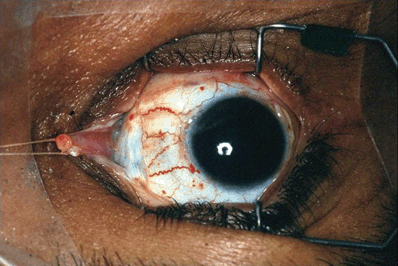
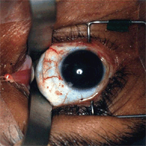
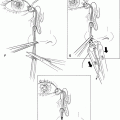
Figure 34-1. The medial approach involves disinsertion of the medial rectus muscle to gain access to the intraconal space and optic nerve. A lid speculum is used to retract the eyelids and a 180° conjunctival peritomy is performed along the medial limbus from 12 to 6 o’clock positions. A tenotomy scissors is used to bluntly dissect along the sclera in the superonasal and inferonasal quadrants. The medial rectus muscle is isolated with a muscle hook and the intramuscular attachments are bluntly separated with a cotton-tipped applicator. A double-armed 5–0 polygalactin suture is passed partial thickness through the medial rectus of the muscle just behind the insertion, with “locking” full-thickness suture bites at the superior and inferior poles of the muscle. The medial rectus muscle is then disinserted with a Westcott scissors and retracted nasally by anchoring the sutures to the drape.
A small branch of the nasociliary nerve extends along the lateral side of the optic nerve and enters the ciliary ganglion. This sensory branch travels through the ganglion without synapsing, and continues as the short posterior ciliary nerves. The long ciliary nerves, two or three in number, extend from the nasociliary nerve as it crosses the optic nerve. These nerves pass by the ciliary ganglion without synapsing and progress with the short posterior ciliary nerves to the globe. These thin, grayish-yellow nerve fibers, oriented along the axis of the optic nerve can be visualized with short posterior ciliary vessels on the epidural surface. Manipulation of these fibers with instruments will cause sectorial pupillary dilation.
The intraconal space between the sclera and the medial rectus is now accessible. Dissection within the central surgical space to gain access to the optic nerve is performed with malleable orbital retractors. Perhaps the most difficult aspect of the dissection is the tendency of orbital fat to obscure normal anatomical landmarks. The fat is divided into lobules by fine connective-tissue septa; these lobules often billow over the edges of the retractors, obscuring the plane of dissection. This problem can be minimized by using dry, 0.5-in. (13-mm) neurosurgical cottonoids to which the orbital fat will adhere slightly, preventing it from billowing over the edges. By gradually removing and reinserting the orbital retractor blades over the cottonoids, the fat can be kept away from the place of dissection. Care should be taken not to traumatize the vortex vein that passes through the orbital fat.
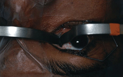
Figure 34-2. Two malleable ribbon retractors (1 cm in width) are bent 90° to form a right angle. The two retractors are used to enter the medial orbit oriented and in alignment with the medial rectus. Once the malleable retractors are in the intraconal space behind the globe, they are rotated 90° so the retractors are oriented superiorly and inferiorly in the respective quadrants. The retractors are then separated to expose the intraconal space with the direction of retraction toward the roof and orbital floor.
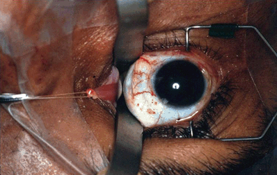
Figure 34-3. When retraction is applied to the blades to separate the intraconal fat, the globe should not rotate or move. If there is globe rotation, the blade of one of the retractors is most likely retracting against the optic nerve, and should be repositioned immediately.
Related posts:
Stay updated, free articles. Join our Telegram channel

Full access? Get Clinical Tree


