United States [45]
Canada [32]
Iran [41]
Brazil [33]
Colombia [34]
Pakistan [35]
Thailand [36]
Italy [37]
Greece [38]
India [4]
Turkey [42]
Total percentage of onychomycosis due to NDM
20.7 %
4.3 %
11.5 %
7.4 %
14 %
11 %
51.6 %
9 %
15.5 %
35.3 %
9 %
Scopulariopsis brevicaulis
20.5 %
–
2.1 %
–
–
18.2 %
–
35.3 %
65.9 %
3.8 %
3 %
Aspergillus species
11.4 %
33 %
59.6 %
8.1 %
–
18.2 %
–
27.5 %
4.5 %
85 %
22 %
Acremonium species
29.5 %
33 %
17 %
–
–
9.1 %
–
5.9 %
22.8 %
–
18 %
Neoscytalidium species
4.5 %
17 %
–
–
38 %
9.1 %
70.6 %
–
–
–
–
Fusarium species
34.1 %
–
12.7 %
60.8 %
42.9 %
36.4 %
29.4 %
27.5 %
–
3.8 %
18 %
Nattrassia mangiferae
–
–
–
31.1 %
–
–
–
–
–
–
–
Alternaria species
–
–
–
–
–
9.1 %
–
–
4.5 %
–
3 %
Ulocladium species
–
–
–
–
–
–
–
–
–
–
12 %
Most cases of mold onychomycosis are caused by Scopulariopsis brevicaulis [3, 11, 20, 37, 38, 40, 43] or Aspergillus spp. [4, 18, 30, 41, 42, 44]. Most infections of non-dermatophytic molds are found in the hot and humid tropical and subtropical parts of the world [3, 21, 30]. Prevalence varies globally, depending on the climate and microenvironment of each geographic region. Acremonium spp., Aspergillus spp., and Neoscytalidium spp. are common in Canada [32]. Fusarium spp., Acremonium spp., and Scopulariopsis brevicaulis are common in the United States [45]. Neoscytalidium spp. and Fusarium spp. are found throughout South America (especially prevalent in Colombia and Brazil) [33, 35]. Scopulariopsis brevicaulis, Aspergillus spp., and Fusarium spp. are frequently isolated in Pakistan [36]. Neoscytalidium dimidiatum and Fusarium spp. are found in Thailand [36], while Scopulariopsis brevicaulis, Aspergillus spp., Acremonium spp., and Fusarium spp. are common in the Mediterranean (Italy, Greece, and Turkey) [37–39, 42].
The most important predisposing factor is, as for dermatophytes, patient’s age. Studies of non-dermatophytic mold onychomycosis report most afflicted patients being older than 40 years [1, 4, 6, 8, 18, 43]. Possible reasons include slower nail growth rate with aging, repeated nail trauma, prolonged exposure to pathogenic fungi, and venous insufficiency. Toenails are generally more often affected than fingernails, due to their slower growth rate.
Onychomycosis from non-dermatophytic molds is more common in females, in contrast to dermatophytic onychomycosis [1, 31, 32]. In a Colombian study of 310 cases of non-dermatophytic mold onychomycosis with toenail infections, women represented 62 % of cases [31].
Clinical Features
Non-dermatophytic molds can cause proximal subungual, “deep” white superficial, and distal subungual onychomycosis.
Proximal subungual onychomycosis is characterized by the invasion of the nail matrix through the proximal nail folds. Fungi are then incorporated in the ventral nail plate from the matrix. The proximal nail plate shows a yellow-white discoloration as presence of fungal elements changes nail plate transparency. Presence of erythema and swelling of proximal and lateral nail folds is common (Fig. 7.1). Inflammation can be prominent in some cases and purulent discharge might occur. Proximal subungual onychomycosis is commonly associated with the following molds: Fusarium spp., Aspergillus spp., and Scopulariopsis brevicaulis [24]. Fusarium and Scopulariopsis brevicaulis produce a yellow-white discoloration of the proximal nail plate [23, 24], while Aspergillus can be associated with a black or green discoloration [25, 26]. The presence of periungual inflammation strongly suggests a mold infection, as this feature is almost never seen in proximal subungual onychomycosis caused by dermatophytes (Fig. 7.2) [5].
Fig. 7.1
Proximal subungual onychomycosis with periungual inflammation due to Fusarium spp.
Fig. 7.2
Proximal subungual onychomycosis with periungual inflammation due to Aspergillus spp.
Deep white superficial onychomycosis is characterized by opaque, friable, white, superficial lesions that start on the dorsal surface of the nail plate, usually on the toes [26]. Mold infections differ from classic white superficial onychomycosis caused by dermatophytes because the infection is deeper and more diffuse (Fig. 7.3a, b) [27, 29]. Deep white superficial onychomycosis is commonly seen with Fusarium spp., Acremonium spp., and Aspergillus spp. [22, 27, 28].
Fig. 7.3
(a, b) Deep white superficial onychomycosis (a) dermoscopy shows invasion of the intermediate nail plate (b)
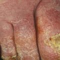
Distal subungual onychomycosis is primarily a nail bed disorder. Infection usually begins with involvement of the distal part of the nail bed and progresses proximally along the ventral surface of the nail plate. It most commonly affects the great toe [7]. The nails become thick due to subungual hyperkeratosis, which is associated with onycholysis. The onycholytic nail plate is yellowish white to brown in color (Fig. 7.4). Agents responsible for distal subungual onychomycosis include Acremonium spp., Fusarium spp., and Alternaria spp. [5, 7, 8]. Periungual inflammation may be seen in distal subungual onychomycosis caused by Fusarium spp. [5]. Tinea pedis is not commonly associated with mold onychomycosis, although it can be seen in Scopulariopsis brevicaulis infections.


Fig. 7.4
Distal subungual onychomycosis from molds
Related posts:

Stay updated, free articles. Join our Telegram channel
Full access? Get Clinical Tree
