Fig. 5.1
Illustration of the classically described mechanism of pathogenesis in PSO. There is invasion of the proximal nail fold and nail matrix by fungi
More recently, alternative routes of infection have been described such as hematogenous spread and spread via the lymphatics, which can be exemplified by maladie dermatophytique [4, 5]. Maladie dermatophytique is a generalized dermatophytosis due to T. schoenleinii that affects the skin and internal organs alike [4]. Severe lymphadenopathy is observed in this condition, and biopsies of these enlarged lymph nodes have shown fungal hyphae, which is evidence of systemic spread of the fungal infection via lymphatics [5]. It is now thought that PSO can also be caused by fungal infection of the bloodstream; however, the exact mechanism of transfer from the blood to the nail plate is unclear [4]. Interestingly, there is some new evidence that PSO can progress from an isolated nail infection to a bloodstream infection, especially in immunosuppressed neutropenic patients [4]. The common link between all proposed mechanisms of infection is that fungi gain access to the proximal matrix that produces the ventral nail plate.
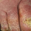
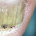
The most common organism causing PSO is Trichophyton rubrum [6], but many other dermatophytes have been identified such as T. schoenleinii [7], T. megnini [6, 8], T. tonsurans [6, 8], T. mentagrophytes [6], T. epidermophyton [6], E. floccosum [6], and Microsporum sp. [9]. Most cases are seen in immunocompromised individuals like HIV/AIDS [6, 8, 10], diabetics, dialysis, and transplant patients [7, 11]. PSO due to dermatophytes is not usually associated with periungual inflammation (Fig. 5.2). Some studies have noted that PSO occurs in up to one third of patients with serious or symptomatic HIV infection and can be considered a sign of immunodeficiency [12]. However, it is important to note that PSO in HIV patients has generally only been studied in those who have progressed to AIDS. Therefore, older data cannot be used to generalize PSO presentation or mycology in all immunosuppressed patients, especially those with higher CD4 counts and without AIDS-defining complications. Moreover, most studies that investigated PSO in AIDS were conducted before the emergence of HAART, which is now the standard of care.
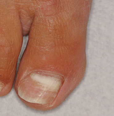
Fig. 5.2
PSO due to T. rubrum seen in an immunosuppressed patient
Several non-dermatophytic molds have been identified as causes of PSO including Fusarium sp. [13, 14], Aspergillus fumigatus [6, 15], Aspergillus flavus [16], Scopulariopsis brevicaulis [17], and Acremonium sp. [17]. Unlike PSO due to dermatophytes, these are not associated with immunosuppressed patients [17]. Non-dermatophytic molds are being increasingly considered a major cause of PSO in otherwise healthy patients. In the original classification of PSO, non-dermatophytic molds were thought to be contaminants, but further investigation has identified them as a significant cause of PSO. Of note, non-dermatophytic PSO is associated with significant periungual inflammation in many cases, which can help distinguish PSO due to dermatophytes from PSO due to non-dermatophytic molds clinically (Fig. 5.3). Distinguishing between dermatophytes and molds is important to guide treatment as their antifungal sensitivities are different.
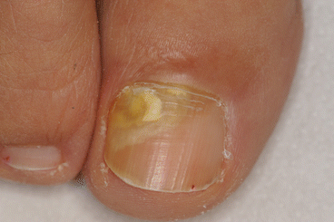
Fig. 5.3
PSO caused by a non-dermatophytic mold
Lastly, PSO can be caused by Candida albicans [6, 18]. Candida PSO is usually associated with paronychia, with the assumption that the inflammation can facilitate yeast invasion of the ventral nail plate. Candida PSO may rarely occur in chronic mucocutaneous candidiasis (CMCC) [18], a disorder of T cells that is characterized by chronic infection of mucosa, skin, and nails by Candida. In CCCA, Candida PSO is not always associated with paronychia and patients have a widespread mucocutaneous infection. Of note, the classic nail appearance in CMCC is granulomatous and totally dystrophic. Although PSO is not the most common nail presentation of CMCC, it can certainly be seen in this condition, and it has been reported in more recent literature [18].
Epidemiology
The exact prevalence of PSO is not clear due to a lack of large studies on the subject. Because it is most prevalent in immunocompromised patients, some studies have investigated PSO in this context. A study of onychomycosis in AIDS patients found that 55 of the 62 patients (88.7 %) who were seen for nail infections presented with PSO [6]. They also found that most (83 %) infections occurred in the feet [6]. Because these patients were immunosuppressed, the most common cause of infection were dermatophytes, with T. rubrum isolated in 36 (58 %) individuals followed by T. mentagrophytes (9.7 %) and Epidermophyton floccosum (4.8 %) [6]. Of note, some yeasts were isolated including Candida albicans (11.2 %) and Pityrosporum ovale (3.2 %) [6]. Of the non-dermatophytic molds, S. brevicaulis and A. fumigatus were isolated in four patients and one patient, respectively [6]. These non-dermatophytes were found coexisting with dermatophytes and never independently. Incorporating these results and other reports of PSO in the literature, PSO due to dermatophytes can be considered a sign of immunodeficiency [12].
Non-dermatophytic molds are another potential cause of PSO and are the most common cause of PSO in patients who are not immunosuppressed. Mold onychomycosis is not significantly associated with systemic diseases and should not be regarded as a sign of immunodeficiency as PSO due to dermatophytes is. A study at the University of Bologna [17] between 1995 and 1998 identified 59 patients out of 1548 who were affected by onychomycosis due to non-dermatophytic molds. Molds were responsible for 13.6 % of all onychomycoses diagnosed via mycology culture, and the majority of these cases (76 %) presented with PSO. Fusarium sp. was identified in 44 % of these cases followed by 29 % Scopulariopsis brevicaulis, 15 % Acremonium sp., and 12 % Aspergillus sp. [17].
Furthermore, this study of non-dermatophytic molds identified an interesting relationship between molds and periungual inflammation. The researchers found that all cases of Fusarium sp. and Aspergillus sp., as well as 59 % of cases of S. brevicaulis, were associated with significant periungual inflammation [17]. This contrasts to classic dermatophyte onychomycoses, which were found to almost never cause inflammation [17]. In fact, the authors noted that many of these patients with inflammation were initially treated with antibiotics and anti-inflammatory drugs due to their clinical presentation [17]. Thus, periungual inflammation is an important factor when developing a differential in PSO, and it may help influence what therapy the clinician will recommend (Fig. 5.4).
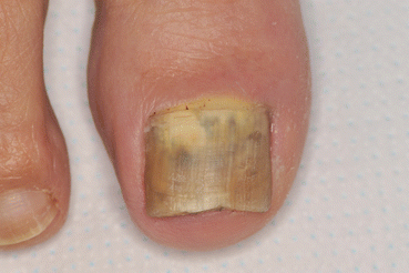
Fig. 5.4
PSO due to mold with associated periungual inflammation
Generally speaking, the exact prevalence of certain fungi in PSO is unclear. In the past, PSO was thought to originate from periungual inflammation of the proximal nail fold. Also, it was commonly believed that non-dermatophytic molds that were isolated in cases of PSO were contaminants and not the cause of infection [17]. However, it has been revealed in more recent literature that PSO due to molds is frequently associated with periungual inflammation and that molds are a more common cause of PSO than was previously thought, which may invalidate much of our historical data. Furthermore, many of the studies analyzing PSO in the presence of HIV infection were conducted before HAART therapy became the standard of care. Many patients who were studied had extremely low CD4 counts, but today, clinicians can expect to see HIV patients with significantly lower degrees of immunosuppression due to HAART. Thus, one must be careful translating older data into modern clinical practice.
Clinical Features
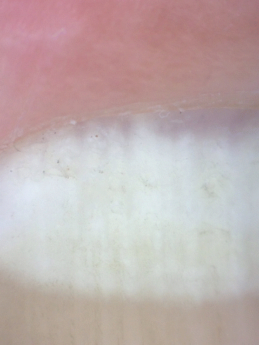
Proximal subungual onychomycosis (PSO) is an infection of the ventral nail plate. It can be seen in both fingers and toes with the toes being more common. In the classic description of PSO, it begins with fungal invasion of the stratum corneum of proximal nail fold. There is subsequent infection of the deeper portions of the ventral nail plate, which is then followed by slow extension of the infection distally as the nail plate grows [1–3]. Eventually, the infection may involve the entire nail plate [1]. Clinically, this infection appears as a white leukonychia spreading distally as the nail plate grows. PSO causes true leukonychia as the white color is due to lack of light reflection due to the presence of fungi within the nail plate (Fig. 5.5). The nail surface is normal. The leukonychia can originate from the proximal nail fold or from the distal matrix with a single band that follows the shape of the lunula (Figs. 5.6 and 5.7).